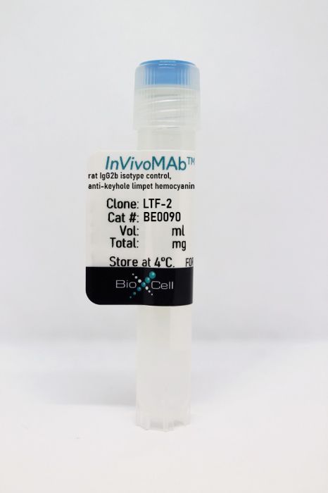
InVivoMAb rat IgG2b isotype control, anti-keyhole limpet hemocyanin
Product Details
The LTF-2 monoclonal antibody reacts with keyhole limpet hemocyanin (KLH). Because KLH is not expressed by mammals this antibody is ideal for use as an isotype-matched control for rat IgG2b antibodies in most in vivo and in vitro applications.Specifications
| Isotype | Rat IgG2b, κ |
|---|---|
| Recommended Dilution Buffer | InVivoPure pH 7.0 Dilution Buffer |
| Conjugation | This product is unconjugated. Conjugation is available via our Antibody Conjugation Services. |
| Formulation |
PBS, pH 7.0 Contains no stabilizers or preservatives |
| Endotoxin |
<2EU/mg (<0.002EU/μg) Determined by LAL gel clotting assay |
| Purity |
>95% Determined by SDS-PAGE |
| Sterility | 0.2 µm filtration |
| Production | Purified from cell culture supernatant in an animal-free facility |
| Purification | Protein G |
| RRID | AB_1107780 |
| Molecular Weight | 150 kDa |
| Storage | The antibody solution should be stored at the stock concentration at 4°C. Do not freeze. |
Additional Formats
Recommended Products
Bauche, D., et al. (2018). "LAG3(+) Regulatory T Cells Restrain Interleukin-23-Producing CX3CR1(+) Gut-Resident Macrophages during Group 3 Innate Lymphoid Cell-Driven Colitis" Immunity 49(2): 342-352 e345. PubMed
Interleukin-22 (IL-22)-producing group 3 innate lymphoid cells (ILC3) maintains gut homeostasis but can also promote inflammatory bowel disease (IBD). The regulation of ILC3-dependent colitis remains to be elucidated. Here we show that Foxp3(+) regulatory T cells (Treg cells) prevented ILC3-mediated colitis in an IL-10-independent manner. Treg cells inhibited IL-23 and IL-1beta production from intestinal-resident CX3CR1(+) macrophages but not CD103(+) dendritic cells. Moreover, Treg cells restrained ILC3 production of IL-22 through suppression of CX3CR1(+) macrophage production of IL-23 and IL-1beta. This suppression was contact dependent and was mediated by latent activation gene-3 (LAG-3)-an immune checkpoint receptor-expressed on Treg cells. Engagement of LAG-3 on MHC class II drove profound immunosuppression of CX3CR1(+) tissue-resident macrophages. Our study reveals that the health of the intestinal mucosa is maintained by an axis driven by Treg cells communication with resident macrophages that withhold inflammatory stimuli required for ILC3 function.
Triplett, T. A., et al. (2018). "Reversal of indoleamine 2,3-dioxygenase-mediated cancer immune suppression by systemic kynurenine depletion with a therapeutic enzyme" Nat Biotechnol 36(8): 758-764. PubMed
Increased tryptophan (Trp) catabolism in the tumor microenvironment (TME) can mediate immune suppression by upregulation of interferon (IFN)-gamma-inducible indoleamine 2,3-dioxygenase (IDO1) and/or ectopic expression of the predominantly liver-restricted enzyme tryptophan 2,3-dioxygenase (TDO). Whether these effects are due to Trp depletion in the TME or mediated by the accumulation of the IDO1 and/or TDO (hereafter referred to as IDO1/TDO) product kynurenine (Kyn) remains controversial. Here we show that administration of a pharmacologically optimized enzyme (PEGylated kynureninase; hereafter referred to as PEG-KYNase) that degrades Kyn into immunologically inert, nontoxic and readily cleared metabolites inhibits tumor growth. Enzyme treatment was associated with a marked increase in the tumor infiltration and proliferation of polyfunctional CD8(+) lymphocytes. We show that PEG-KYNase administration had substantial therapeutic effects when combined with approved checkpoint inhibitors or with a cancer vaccine for the treatment of large B16-F10 melanoma, 4T1 breast carcinoma or CT26 colon carcinoma tumors. PEG-KYNase mediated prolonged depletion of Kyn in the TME and reversed the modulatory effects of IDO1/TDO upregulation in the TME.
Aloulou, M., et al. (2016). "Follicular regulatory T cells can be specific for the immunizing antigen and derive from naive T cells" Nat Commun 7: 10579. PubMed
T follicular regulatory (Tfr) cells are a subset of Foxp3(+) regulatory T (Treg) cells that form in response to immunization or infection, which localize to the germinal centre where they control the magnitude of the response. Despite an increased interest in the role of Tfr cells in humoral immunity, many fundamental aspects of their biology remain unknown, including whether they recognize self- or foreign antigen. Here we show that Tfr cells can be specific for the immunizing antigen, irrespective of whether it is a self- or foreign antigen. We show that, in addition to developing from thymic derived Treg cells, Tfr cells can also arise from Foxp3(-) precursors in a PD-L1-dependent manner, if the adjuvant used is one that supports T-cell plasticity. These findings have important implications for Tfr cell biology and for improving vaccine efficacy by formulating vaccines that modify the Tfr:Tfh cell ratio.
Finkin, S., et al. (2015). "Ectopic lymphoid structures function as microniches for tumor progenitor cells in hepatocellular carcinoma" Nat Immunol. doi : 10.1038/ni.3290. PubMed
Ectopic lymphoid-like structures (ELSs) are often observed in cancer, yet their function is obscure. Although ELSs signify good prognosis in certain malignancies, we found that hepatic ELSs indicated poor prognosis for hepatocellular carcinoma (HCC). We studied an HCC mouse model that displayed abundant ELSs and found that they constituted immunopathological microniches wherein malignant hepatocyte progenitor cells appeared and thrived in a complex cellular and cytokine milieu until gaining self-sufficiency. The egress of progenitor cells and tumor formation were associated with the autocrine production of cytokines previously provided by the niche. ELSs developed via cooperation between the innate immune system and adaptive immune system, an event facilitated by activation of the transcription factor NF-kappaB and abolished by depletion of T cells. Such aberrant immunological foci might represent new targets for cancer therapy.
Zhang, J., et al. (2015). "Micro-RNA-155-mediated control of heme oxygenase 1 (HO-1) is required for restoring adaptively tolerant CD4+ T-cell function in rodents" Eur J Immunol 45(3): 829-842. PubMed
T cells chronically stimulated by a persistent antigen often become dysfunctional and lose effector functions and proliferative capacity. To identify the importance of micro-RNA-155 (miR-155) in this phenomenon, we analyzed mouse miR-155-deficient CD4(+) T cells in a model where the chronic exposure to a systemic antigen led to T-cell functional unresponsiveness. We found that miR-155 was required for restoring function of T cells after programmed death receptor 1 blockade. Heme oxygenase 1 (HO-1) was identified as a specific target of miR-155 and inhibition of HO-1 activity restored the expansion and tissue migration capacity of miR-155(-/-) CD4(+) T cells. Moreover, miR-155-mediated control of HO-1 expression in CD4(+) T cells was shown to sustain in vivo antigen-specific expansion and IL-2 production. Thus, our data identify HO-1 regulation as a mechanism by which miR-155 promotes T-cell-driven inflammation.
Park, H. J., et al. (2015). "PD-1 upregulated on regulatory T cells during chronic virus infection enhances the suppression of CD8+ T cell immune response via the interaction with PD-L1 expressed on CD8+ T cells" J Immunol 194(12): 5801-5811. PubMed
Regulatory T (Treg) cells act as terminators of T cell immuniy during acute phase of viral infection; however, their role and suppressive mechanism in chronic viral infection are not completely understood. In this study, we compared the phenotype and function of Treg cells during acute or chronic infection with lymphocytic choriomeningitis virus. Chronic infection, unlike acute infection, led to a large expansion of Treg cells and their upregulation of programmed death-1 (PD-1). Treg cells from chronically infected mice (chronic Treg cells) displayed greater suppressive capacity for inhibiting both CD8(+) and CD4(+) T cell proliferation and subsequent cytokine production than those from naive or acutely infected mice. A contact between Treg and CD8(+) T cells was necessary for the potent suppression of CD8(+) T cell immune response. More importantly, the suppression required cell-specific expression and interaction of PD-1 on chronic Treg cells and PD-1 ligand on CD8(+) T cells. Our study defines PD-1 upregulated on Treg cells and its interaction with PD-1 ligand on effector T cells as one cause for the potent T cell suppression and proposes the role of PD-1 on Treg cells, in addition to that on exhausted T cells, during chronic viral infection.
Twyman-Saint Victor, C., et al. (2015). "Radiation and dual checkpoint blockade activate non-redundant immune mechanisms in cancer" Nature 520(7547): 373-377. PubMed
Immune checkpoint inhibitors result in impressive clinical responses, but optimal results will require combination with each other and other therapies. This raises fundamental questions about mechanisms of non-redundancy and resistance. Here we report major tumour regressions in a subset of patients with metastatic melanoma treated with an anti-CTLA4 antibody (anti-CTLA4) and radiation, and reproduced this effect in mouse models. Although combined treatment improved responses in irradiated and unirradiated tumours, resistance was common. Unbiased analyses of mice revealed that resistance was due to upregulation of PD-L1 on melanoma cells and associated with T-cell exhaustion. Accordingly, optimal response in melanoma and other cancer types requires radiation, anti-CTLA4 and anti-PD-L1/PD-1. Anti-CTLA4 predominantly inhibits T-regulatory cells (Treg cells), thereby increasing the CD8 T-cell to Treg (CD8/Treg) ratio. Radiation enhances the diversity of the T-cell receptor (TCR) repertoire of intratumoral T cells. Together, anti-CTLA4 promotes expansion of T cells, while radiation shapes the TCR repertoire of the expanded peripheral clones. Addition of PD-L1 blockade reverses T-cell exhaustion to mitigate depression in the CD8/Treg ratio and further encourages oligoclonal T-cell expansion. Similarly to results from mice, patients on our clinical trial with melanoma showing high PD-L1 did not respond to radiation plus anti-CTLA4, demonstrated persistent T-cell exhaustion, and rapidly progressed. Thus, PD-L1 on melanoma cells allows tumours to escape anti-CTLA4-based therapy, and the combination of radiation, anti-CTLA4 and anti-PD-L1 promotes response and immunity through distinct mechanisms.
Rutigliano, J. A., et al. (2014). "Highly pathological influenza A virus infection is associated with augmented expression of PD-1 by functionally compromised virus-specific CD8+ T cells" J Virol 88(3): 1636-1651. PubMed
One question that continues to challenge influenza A research is why some strains of virus are so devastating compared to their more mild counterparts. We approached this question from an immunological perspective, investigating the CD8(+) T cell response in a mouse model system comparing high- and low-pathological influenza virus infections. Our findings reveal that the early (day 0 to 5) viral titer was not the determining factor in the outcome of disease. Instead, increased numbers of antigen-specific CD8(+) T cells and elevated effector function on a per-cell basis were found in the low-pathological infection and correlated with reduced illness and later-time-point (day 6 to 10) viral titer. High-pathological infection was associated with increased PD-1 expression on influenza virus-specific CD8(+) T cells, and blockade of PD-L1 in vivo led to reduced virus titers and increased CD8(+) T cell numbers in high- but not low-pathological infection, though T cell functionality was not restored. These data show that high-pathological acute influenza virus infection is associated with a dysregulated CD8(+) T cell response, which is likely caused by the more highly inflamed airway microenvironment during the early days of infection. Therapeutic approaches specifically aimed at modulating innate airway inflammation may therefore promote efficient CD8(+) T cell activity. We show that during a severe influenza virus infection, one type of immune cell, the CD8 T cell, is less abundant and less functional than in a more mild infection. This dysregulated T cell phenotype correlates with a lower rate of virus clearance in the severe infection and is partially regulated by the expression of a suppressive coreceptor called PD-1. Treatment with an antibody that blocks PD-1 improves T cell functionality and increases virus clearance.
Erickson, J. J., et al. (2014). "Programmed death-1 impairs secondary effector lung CD8(+) T cells during respiratory virus reinfection" J Immunol 193(10): 5108-5117. PubMed
Reinfections with respiratory viruses are common and cause significant clinical illness, yet precise mechanisms governing this susceptibility are ill defined. Lung Ag-specific CD8(+) T cells (T(CD8)) are impaired during acute viral lower respiratory infection by the inhibitory receptor programmed death-1 (PD-1). To determine whether PD-1 contributes to recurrent infection, we first established a model of reinfection by challenging B cell-deficient mice with human metapneumovirus (HMPV) several weeks after primary infection, and found that HMPV replicated to high titers in the lungs. A robust secondary effector lung TCD8 response was generated during reinfection, but these cells were more impaired and more highly expressed the inhibitory receptors PD-1, LAG-3, and 2B4 than primary T(CD8). In vitro blockade demonstrated that PD-1 was the dominant inhibitory receptor early after reinfection. In vivo therapeutic PD-1 blockade during HMPV reinfection restored lung T(CD8) effector functions (i.e., degranulation and cytokine production) and enhanced viral clearance. PD-1 also limited the protective efficacy of HMPV epitope-specific peptide vaccination and impaired lung T(CD8) during heterotypic influenza virus challenge infection. Our results indicate that PD-1 signaling may contribute to respiratory virus reinfection and evasion of vaccine-elicited immune responses. These results have important implications for the design of effective vaccines against respiratory viruses.
Steel, C. D., et al. (2014). "Role of peripheral immune response in microglia activation and regulation of brain chemokine and proinflammatory cytokine responses induced during VSV encephalitis" J Neuroimmunol 267(1-2): 50-60. PubMed
We report herein that neuroinvasion by vesicular stomatitis virus (VSV) activates microglia and induces a peripheral dendritic cell (DC)-dependent inflammatory response in the central nervous system (CNS). VSV neuroinvasion rapidly induces multiple brain chemokine and proinflammatory cytokine mRNAs that display bimodal kinetics. Peripheral DC ablation or T cell depletion suppresses the second wave of this response demonstrating that infiltrating T cells are primarily responsible for the bimodal characteristics of this response. The robust infiltrate associated with VSV encephalitis likely depends on sustained production of brain CCL19 and CCR7 expression on infiltrating inflammatory cells.
Kearl, T. J., et al. (2013). "Programmed death receptor-1/programmed death receptor ligand-1 blockade after transient lymphodepletion to treat myeloma" J Immunol 190(11): 5620-5628. PubMed
Early phase clinical trials targeting the programmed death receptor-1/ligand-1 (PD-1/PD-L1) pathway to overcome tumor-mediated immunosuppression have reported promising results for a variety of cancers. This pathway appears to play an important role in the failure of immune reactivity to malignant plasma cells in multiple myeloma patients, as the tumor cells express relatively high levels of PD-L1, and T cells show increased PD-1 expression. In the current study, we demonstrate that PD-1/PD-L1 blockade with a PD-L1-specific Ab elicits rejection of a murine myeloma when combined with lymphodepleting irradiation. This particular combined approach by itself has not previously been shown to be efficacious in other tumor models. The antitumor effect of lymphodepletion/anti-PD-L1 therapy was most robust when tumor Ag-experienced T cells were present either through cell transfer or survival after nonmyeloablative irradiation. In vivo depletion of CD4 or CD8 T cells completely eliminated antitumor efficacy of the lymphodepletion/anti-PD-L1 therapy, indicating that both T cell subsets are necessary for tumor rejection. Elimination of myeloma by T cells occurs relatively quickly as tumor cells in the bone marrow were nearly nondetectable by 5 d after the first anti-PD-L1 treatment, suggesting that antimyeloma reactivity is primarily mediated by preactivated T cells, rather than newly generated myeloma-reactive T cells. Anti-PD-L1 plus lymphodepletion failed to improve survival in two solid tumor models, but demonstrated significant efficacy in two hematologic malignancy models. In summary, our results support the clinical testing of lymphodepletion and PD-1/PD-L1 blockade as a novel approach for improving the survival of patients with multiple myeloma.
van der Merwe, M., et al. (2013). "Recipient myeloid-derived immunomodulatory cells induce PD-1 ligand-dependent donor CD4+Foxp3+ regulatory T cell proliferation and donor-recipient immune tolerance after murine nonmyeloablative bone marrow transplantation" J Immunol 191(11): 5764-5776. PubMed
We showed previously that nonmyeloablative total lymphoid irradiation/rabbit anti-thymocyte serum (TLI/ATS) conditioning facilitates potent donor-recipient immune tolerance following bone marrow transplantation (BMT) across MHC barriers via recipient invariant NKT (iNKT) cell-derived IL-4-dependent expansion of donor Foxp3(+) naturally occurring regulatory T cells (nTregs). In this study, we report a more specific mechanism. Wild-type (WT) BALB/c (H-2(d)) hosts were administered TLI/ATS and BMT from WT or STAT6(-/-) C57BL/6 (H-2(b)) donors. Following STAT6(-/-) BMT, donor nTregs demonstrated no loss of proliferation in vivo, indicating that an IL-4-responsive population in the recipient, rather than the donor, drives donor nTreg proliferation. In graft-versus-host disease (GVHD) target organs, three recipient CD11b(+) cell subsets (Gr-1(high)CD11c(-), Gr-1(int)CD11c(-), and Gr-1(low)CD11c(+)) were enriched early after TLI/ATS + BMT versus total body irradiation/ATS + BMT. Gr-1(low)CD11c(+) cells induced potent H-2K(b+)CD4(+)Foxp3(+) nTreg proliferation in vitro in 72-h MLRs. Gr-1(low)CD11c(+) cells were reduced significantly in STAT6(-/-) and iNKT cell-deficient Jalpha18(-/-) BALB/c recipients after TLI/ATS + BMT. Depletion of CD11b(+) cells resulted in severe acute GVHD, and adoptive transfer of WT Gr-1(low)CD11c(+) cells to Jalpha18(-/-) BALB/c recipients of TLI/ATS + BMT restored day-6 donor Foxp3(+) nTreg proliferation and protection from CD8 effector T cell-mediated GVHD. Blockade of programmed death ligand 1 and 2, but not CD40, TGF-beta signaling, arginase 1, or iNOS, inhibited nTreg proliferation in cocultures of recipient-derived Gr-1(low)CD11c(+) cells with donor nTregs. Through iNKT-dependent Th2 polarization, myeloid-derived immunomodulatory dendritic cells are expanded after nonmyeloablative TLI/ATS conditioning and allogeneic BMT, induce PD-1 ligand-dependent donor nTreg proliferation, and maintain potent graft-versus-host immune tolerance.
Sledzinska, A., et al. (2013). "TGF-beta signalling is required for CD4(+) T cell homeostasis but dispensable for regulatory T cell function" PLoS Biol 11(10): e1001674. PubMed
TGF-beta is widely held to be critical for the maintenance and function of regulatory T (T(reg)) cells and thus peripheral tolerance. This is highlighted by constitutive ablation of TGF-beta receptor (TR) during thymic development in mice, which leads to a lethal autoimmune syndrome. Here we describe that TGF-beta-driven peripheral tolerance is not regulated by TGF-beta signalling on mature CD4(+) T cells. Inducible TR2 ablation specifically on CD4(+) T cells did not result in a lethal autoinflammation. Transfer of these TR2-deficient CD4(+) T cells to lymphopenic recipients resulted in colitis, but not overt autoimmunity. In contrast, thymic ablation of TR2 in combination with lymphopenia led to lethal multi-organ inflammation. Interestingly, deletion of TR2 on mature CD4(+) T cells does not result in the collapse of the T(reg) cell population as observed in constitutive models. Instead, a pronounced enlargement of both regulatory and effector memory T cell pools was observed. This expansion is cell-intrinsic and seems to be caused by increased T cell receptor sensitivity independently of common gamma chain-dependent cytokine signals. The expression of Foxp3 and other regulatory T cells markers was not dependent on TGF-beta signalling and the TR2-deficient T(reg) cells retained their suppressive function both in vitro and in vivo. In summary, absence of TGF-beta signalling on mature CD4(+) T cells is not responsible for breakdown of peripheral tolerance, but rather controls homeostasis of mature T cells in adult mice.
Willimsky, G., et al. (2013). "Virus-induced hepatocellular carcinomas cause antigen-specific local tolerance" J Clin Invest 123(3): 1032-1043. PubMed
T cell surveillance is often effective against virus-associated tumors because of their high immunogenicity. It is not clear why surveillance occasionally fails, particularly against hepatitis B virus- or hepatitis C virus-associated hepatocellular carcinoma (HCC). We established a transgenic murine model of virus-induced HCC by hepatocyte-specific adenovirus-induced activation of the oncogenic SV40 large T antigen (TAg). Adenovirus infection induced cytotoxic T lymphocytes (CTLs) targeted against the virus and TAg, leading to clearance of the infected cells. Despite the presence of functional, antigen-specific T cells, a few virus-infected cells escaped immune clearance and progressed to HCC. These cells expressed TAg at levels similar to HCC isolated from neonatal TAg-tolerant mice, suggesting that CTL clearance does not select for cells with low immunogenicity. Virus-infected mice revealed significantly greater T cell infiltration in early-stage HCC compared with that in late-stage HCC, demonstrating progressive local immune suppression through inefficient T cell infiltration. Programmed cell death protein-1 (PD-1) and its ligand PD-L1 were expressed in all TAg-specific CD8+ T cells and HCC, respectively, which contributed to local tumor-antigen-specific tolerance. Thus, we have developed a model of virus-induced HCC that may allow for a better understanding of human HCC.
Coers, J., et al. (2011). "Compensatory T cell responses in IRG-deficient mice prevent sustained Chlamydia trachomatis infections" PLoS Pathog 7(6): e1001346. PubMed
The obligate intracellular pathogen Chlamydia trachomatis is the most common cause of bacterial sexually transmitted diseases in the United States. In women C. trachomatis can establish persistent genital infections that lead to pelvic inflammatory disease and sterility. In contrast to natural infections in humans, experimentally induced infections with C. trachomatis in mice are rapidly cleared. The cytokine interferon-gamma (IFNgamma) plays a critical role in the clearance of C. trachomatis infections in mice. Because IFNgamma induces an antimicrobial defense system in mice but not in humans that is composed of a large family of Immunity Related GTPases (IRGs), we questioned whether mice deficient in IRG immunity would develop persistent infections with C. trachomatis as observed in human patients. We found that IRG-deficient Irgm1/m3((-/-)) mice transiently develop high bacterial burden post intrauterine infection, but subsequently clear the infection more efficiently than wildtype mice. We show that the delayed but highly effective clearance of intrauterine C. trachomatis infections in Irgm1/m3((-/-)) mice is dependent on an exacerbated CD4(+) T cell response. These findings indicate that the absence of the predominant murine innate effector mechanism restricting C. trachomatis growth inside epithelial cells results in a compensatory adaptive immune response, which is at least in part driven by CD4(+) T cells and prevents the establishment of a persistent infection in mice.

- Immunology and Microbiology,
- Pathology
CD8+ T Cells Mediate Lethal Lung Pathology in the Absence of PD-L1 and Type I Interferon Signalling following LCMV Infection.
In Viruses on 1 March 2024 by Spiteri, A. G., Suprunenko, T., et al.
PubMed
CD8+ T cells are critical to the adaptive immune response against viral pathogens. However, overwhelming antigen exposure can result in their exhaustion, characterised by reduced effector function, failure to clear virus, and the upregulation of inhibitory receptors, including programmed cell death 1 (PD-1). However, exhausted T cell responses can be "re-invigorated" by inhibiting PD-1 or the primary ligand of PD-1: PD-L1. Further, the absence of the type I interferon receptor IFNAR1 also results in T cell exhaustion and virus persistence in lymphocytic choriomeningitis virus Armstrong (LCMV-Arm)-infected mice. In this study, utilizing single- and double-knockout mice, we aimed to determine whether ablation of PD-1 could restore T cell functionality in the absence of IFNAR1 signalling in LCMV-Arm-infected mice. Surprisingly, this did not re-invigorate the T cell response and instead, it converted chronic LCMV-Arm infection into a lethal disease characterized by severe lung inflammation with an infiltration of neutrophils and T cells. Depletion of CD8+ T cells, but not neutrophils, rescued mice from lethal disease, demonstrating that IFNAR1 is required to prevent T cell exhaustion and virus persistence in LCMV-Arm infection, and in the absence of IFNAR1, PD-L1 is required for survival. This reveals an important interplay between IFNAR1 and PD-L1 with implications for therapeutics targeting these pathways.
- Immunology and Microbiology
A viral-specific CD4+ T cell response protects female mice from Coxsackievirus B3 infection.
In Frontiers in Immunology on 26 January 2024 by Pattnaik, A., Dhalech, A. H., et al.
PubMed
Biological sex plays an integral role in the immune response to various pathogens. The underlying basis for these sex differences is still not well defined. Here, we show that Coxsackievirus B3 (CVB3) induces a viral-specific CD4+ T cell response that can protect female mice from mortality. We inoculated C57BL/6 Ifnar-/- mice with CVB3. We investigated the T cell response in the spleen and mesenteric lymph nodes in male and female mice following infection. We found that CVB3 can induce expansion of CD62Llo CD4+ T cells in the mesenteric lymph node and spleen of female but not male mice as early as 5 days post-inoculation, indicative of activation. Using a recombinant CVB3 virus expressing a model CD4+ T cell epitope, we found that this response is due to viral antigen and not bystander activation. Finally, the depletion of CD4+ T cells before infection increased mortality in female mice, indicating that CD4+ T cells play a protective role against CVB3 in our model. Overall, these data demonstrated that CVB3 can induce an early CD4 response in female but not male mice and further emphasize how sex differences in immune responses to pathogens affect disease. Copyright © 2024 Pattnaik, Dhalech, Condotta, Corn, Richer, Snell and Robinson.
- Cancer Research,
- Immunology and Microbiology
Interplay between ATRX and IDH1 mutations governs innate immune responses in diffuse gliomas.
In Nature Communications on 25 January 2024 by Hariharan, S., Whitfield, B. T., et al.
PubMed
Stimulating the innate immune system has been explored as a therapeutic option for the treatment of gliomas. Inactivating mutations in ATRX, defining molecular alterations in IDH-mutant astrocytomas, have been implicated in dysfunctional immune signaling. However, little is known about the interplay between ATRX loss and IDH mutation on innate immunity. To explore this, we generated ATRX-deficient glioma models in the presence and absence of the IDH1R132H mutation. ATRX-deficient glioma cells are sensitive to dsRNA-based innate immune agonism and exhibit impaired lethality and increased T-cell infiltration in vivo. However, the presence of IDH1R132H dampens baseline expression of key innate immune genes and cytokines in a manner restored by genetic and pharmacological IDH1R132H inhibition. IDH1R132H co-expression does not interfere with the ATRX deficiency-mediated sensitivity to dsRNA. Thus, ATRX loss primes cells for recognition of dsRNA, while IDH1R132H reversibly masks this priming. This work reveals innate immunity as a therapeutic vulnerability of astrocytomas. © 2024. The Author(s).
- COVID-19,
- Immunology and Microbiology,
- Pathology
Adaptive immune cells are necessary for SARS-CoV-2-induced pathology.
In Science Advances on 5 January 2024 by Imbiakha, B., Sahler, J., et al.
PubMed
Severe acute respiratory syndrome coronavirus 2 (SARS-CoV-2) is causing the ongoing global pandemic associated with morbidity and mortality in humans. Although disease severity correlates with immune dysregulation, the cellular mechanisms of inflammation and pathogenesis of COVID-19 remain relatively poorly understood. Here, we used mouse-adapted SARS-CoV-2 strain MA10 to investigate the role of adaptive immune cells in disease. We found that while infected wild-type mice lost ~10% weight by 3 to 4 days postinfection, rag-/- mice lacking B and T lymphocytes did not lose weight. Infected lungs at peak weight loss revealed lower pathology scores, fewer neutrophils, and lower interleukin-6 and tumor necrosis factor-α in rag-/- mice. Mice lacking αβ T cells also had less severe weight loss, but adoptive transfer of T and B cells into rag-/- mice did not significantly change the response. Collectively, these findings suggest that while adaptive immune cells are important for clearing SARS-CoV-2 infection, this comes at the expense of increased inflammation and pathology.
- Immunology and Microbiology,
- Cancer Research
Pharmaceutical targeting of OTUB2 sensitizes tumors to cytotoxic T cells via degradation of PD-L1.
In Nature Communications on 2 January 2024 by Ren, W., Xu, Z., et al.
PubMed
PD-1 is a co-inhibitory receptor expressed by CD8+ T cells which limits their cytotoxicity. PD-L1 expression on cancer cells contributes to immune evasion by cancers, thus, understanding the mechanisms that regulate PD-L1 protein levels in cancers is important. Here we identify tumor-cell-expressed otubain-2 (OTUB2) as a negative regulator of antitumor immunity, acting through the PD-1/PD-L1 axis in various human cancers. Mechanistically, OTUB2 directly interacts with PD-L1 to disrupt the ubiquitination and degradation of PD-L1 in the endoplasmic reticulum. Genetic deletion of OTUB2 markedly decreases the expression of PD-L1 proteins on the tumor cell surface, resulting in increased tumor cell sensitivity to CD8+ T-cell-mediated cytotoxicity. To underscore relevance in human patients, we observe a significant correlation between OTUB2 expression and PD-L1 abundance in human non-small cell lung cancer. An inhibitor of OTUB2, interfering with its deubiquitinase activity without disrupting the OTUB2-PD-L1 interaction, successfully reduces PD-L1 expression in tumor cells and suppressed tumor growth. Together, these results reveal the roles of OTUB2 in PD-L1 regulation and tumor evasion and lays down the proof of principle for OTUB2 targeting as therapeutic strategy for cancer treatment. © 2024. The Author(s).
- Immunology and Microbiology
A partial human LCK defect causes a T cell immunodeficiency with intestinal inflammation.
In The Journal of Experimental Medicine on 1 January 2024 by Lui, V. G., Hoenig, M., et al.
PubMed
Lymphocyte-specific protein tyrosine kinase (LCK) is essential for T cell antigen receptor (TCR)-mediated signal transduction. Here, we report two siblings homozygous for a novel LCK variant (c.1318C>T; P440S) characterized by T cell lymphopenia with skewed memory phenotype, infant-onset recurrent infections, failure to thrive, and protracted diarrhea. The patients' T cells show residual TCR signal transduction and proliferation following anti-CD3/CD28 and phytohemagglutinin (PHA) stimulation. We demonstrate in mouse models that complete (Lck-/-) versus partial (LckP440S/P440S) loss-of-function LCK causes disease with differing phenotypes. While both Lck-/- and LckP440S/P440S mice exhibit arrested thymic T cell development and profound T cell lymphopenia, only LckP440S/P440S mice show residual T cell proliferation, cytokine production, and intestinal inflammation. Furthermore, the intestinal disease in the LckP440S/P440S mice is prevented by CD4+ T cell depletion or regulatory T cell transfer. These findings demonstrate that P440S LCK spares sufficient T cell function to allow the maturation of some conventional T cells but not regulatory T cells-leading to intestinal inflammation. © 2023 Lui et al.
- Cancer Research,
- Immunology and Microbiology
Helicobacter pylori CagA promotes immune evasion of gastric cancer by upregulating PD-L1 level in exosomes.
In IScience on 15 December 2023 by Wang, J., Deng, R., et al.
PubMed
Cytotoxin-associated gene A (CagA) of Helicobacter pylori (Hp) may promote immune evasion of Hp-infected gastric cancer (GC), but potential mechanisms are still under explored. In this study, the positive rates of CagA and PD-L1 protein in tumor tissues and the high level of exosomal PD-L1 protein in plasma exosomes were significantly associated with the elevated stages of tumor node metastasis (TNM) in Hp-infected GC. Moreover, the positive rate of CagA was positively correlated with the positive rate of PD-L1 in tumor tissues and the level of PD-L1 protein in plasma exosomes, and high level of exosomal PD-L1 might indicate poor prognosis of Hp-infected GC. Mechanically, CagA increased PD-L1 level in exosomes derived from GC cells by inhibiting p53 and miRNA-34a, suppressing proliferation and anticancer effect of CD8+ T cells. This study provides sights for understanding immune evasion mediated by PD-L1. Targeting CagA and exosomal PD-L1 may improve immunotherapy efficacy of Hp-infected GC. © 2023 The Authors.
High-titer AAV disrupts cerebrovascular integrity and induces lymphocyte infiltration in adult mouse brain.
In Molecular Therapy. Methods Clinical Development on 14 December 2023 by Guo, Y., Chen, J., et al.
PubMed
The brain is often described as an "immune-privileged" organ due to the presence of the blood-brain-barrier (BBB), which limits the entry of immune cells. In general, intracranial injection of adeno-associated virus (AAV) is considered a relatively safe procedure. In this study, we discovered that AAV, a popular engineered viral vector for gene therapy, can disrupt the BBB and induce immune cell infiltration in a titer-dependent manner. First, our bulk RNA sequencing data revealed that injection of high-titer AAV significantly upregulated many genes involved in disrupting BBB integrity and antiviral adaptive immune responses. By using histologic analysis, we further demonstrated that the biological structure of the BBB was severely disrupted in the adult mouse brain. Meanwhile, we noticed abnormal leakage of blood components, including immune cells, within the brain parenchyma of high-titer AAV injected areas. Moreover, we identified that the majority of infiltrated immune cells were cytotoxic T lymphocytes (CTLs), which resulted in a massive loss of neurons at the site of AAV injection. In addition, antagonizing CTL function by administering antibodies significantly reduced neuronal toxicity induced by high-titer AAV. Collectively, our findings underscore potential severe side effects of intracranial injection of high-titer AAV, which might compromise proper data interpretation if unaware of. © 2023 The Authors.
- Cancer Research,
- Immunology and Microbiology
Inhibition of ACLY overcomes cancer immunotherapy resistance via polyunsaturated fatty acids peroxidation and cGAS-STING activation.
In Science Advances on 8 December 2023 by Xiang, W., Lv, H., et al.
PubMed
Adenosine 5'-triphosphate citrate lyase (ACLY) is a cytosolic enzyme that converts citrate into acetyl-coenzyme A for fatty acid and cholesterol biosynthesis. ACLY is up-regulated or activated in many cancers, and targeting ACLY by inhibitors holds promise as potential cancer therapy. However, the role of ACLY in cancer immunity regulation remains poorly understood. Here, we show that ACLY inhibition up-regulates PD-L1 immune checkpoint expression in cancer cells and induces T cell dysfunction to drive immunosuppression and compromise its antitumor effect in immunocompetent mice. Mechanistically, ACLY inhibition causes polyunsaturated fatty acid (PUFA) peroxidation and mitochondrial damage, which triggers mitochondrial DNA leakage to activate the cGAS-STING innate immune pathway. Pharmacological and genetic inhibition of ACLY overcomes cancer resistance to anti-PD-L1 therapy in a cGAS-dependent manner. Furthermore, dietary PUFA supplementation mirrors the enhanced efficacy of PD-L1 blockade by ACLY inhibition. These findings reveal an immunomodulatory role of ACLY and provide combinatorial strategies to overcome immunotherapy resistance in tumors.
- Mus musculus (House mouse),
- Cancer Research,
- Immunology and Microbiology
CD4 T cell-activating neoantigens enhance personalized cancer vaccine efficacy.
In JCI Insight on 8 December 2023 by Huff, A. L., Longway, G., et al.
PubMed
Personalized cancer vaccines aim to activate and expand cytotoxic antitumor CD8+ T cells to recognize and kill tumor cells. However, the role of CD4+ T cell activation in the clinical benefit of these vaccines is not well defined. We previously established a personalized neoantigen vaccine (PancVAX) for the pancreatic cancer cell line Panc02, which activates tumor-specific CD8+ T cells but required combinatorial checkpoint modulators to achieve therapeutic efficacy. To determine the effects of neoantigen-specific CD4+ T cell activation, we generated a vaccine (PancVAX2) targeting both major histocompatibility complex class I- (MHCI-) and MHCII-specific neoantigens. Tumor-bearing mice vaccinated with PancVAX2 had significantly improved control of tumor growth and long-term survival benefit without concurrent administration of checkpoint inhibitors. PancVAX2 significantly enhanced priming and recruitment of neoantigen-specific CD8+ T cells into the tumor with lower PD-1 expression after reactivation compared with the CD8+ vaccine alone. Vaccine-induced neoantigen-specific Th1 CD4+ T cells in the tumor were associated with decreased Tregs. Consistent with this, PancVAX2 was associated with more proimmune myeloid-derived suppressor cells and M1-like macrophages in the tumor, demonstrating a less immunosuppressive tumor microenvironment. This study demonstrates the biological importance of prioritizing and including CD4+ T cell-specific neoantigens for personalized cancer vaccine modalities.
- Cancer Research,
- Cell Biology,
- Immunology and Microbiology
Inhibition of SF3B1 improves the immune microenvironment through pyroptosis and synergizes with αPDL1 in ovarian cancer.
In Cell Death & Disease on 27 November 2023 by Wang, S., Liu, Y., et al.
PubMed
Ovarian cancer is resistant to immune checkpoint blockade (ICB) treatment. Combination of targeted therapy and immunotherapy is a promising strategy for ovarian cancer treatment benefit from an improved immune microenvironment. In this study, Clinical Proteomic Tumor Analysis Consortium (CPTAC) and The Cancer Genome Atlas (TCGA) cohorts were used to screen prognosis and cytotoxic lymphocyte infiltration-associated genes in upregulated genes of ovarian cancer, tissue microarrays were built for further verification. In vitro experiments and mouse (C57/BL6) ovarian tumor (ID8) models were built to evaluate the synergistic effect of the combination of SF3B1 inhibitor and PD-L1 antibody in the treatment of ovarian cancer. The results show that SF3B1 is shown to be overexpressed and related to low cytotoxic immune cell infiltration in ovarian cancer. Inhibition of SF3B1 induces pyroptosis in ovarian cancer cells and releases mitochondrial DNA (mtDNA), which is englobed by macrophages and subsequently activates them (polarization to M1). Moreover, pladienolide B increases cytotoxic immune cell infiltration in the ID8 mouse model as a SF3B1 inhibitor and increases the expression of PD-L1 which can enhance the antitumor effect of αPDL1 in ovarian cancer. The data suggests that inhibition of SF3B1 improves the immune microenvironment of ovarian cancer and synergizes ICB immunotherapy, which provides preclinical evidence for the combination of SF3B1 inhibitor and ICB to ovarian cancer treatment. © 2023. The Author(s).
- Cancer Research,
- Immunology and Microbiology
Targeted Inhibition of lncRNA Malat1 Alters the Tumor Immune Microenvironment in Preclinical Syngeneic Mouse Models of Triple-Negative Breast Cancer.
In Cancer Immunology Research on 1 November 2023 by Oluwatoyosi, A., Shen, Y., et al.
PubMed
Long noncoding RNAs (lncRNA) play an important role in gene regulation in both normal tissues and cancer. Targeting lncRNAs is a promising therapeutic approach that has become feasible through the development of gapmer antisense oligonucleotides (ASO). Metastasis-associated lung adenocarcinoma transcript (Malat1) is an abundant lncRNA whose expression is upregulated in several cancers. Although Malat1 increases the migratory and invasive properties of tumor cells, its role in the tumor microenvironment (TME) is still not well defined. We explored the connection between Malat1 and the tumor immune microenvironment (TIME) using several immune-competent preclinical syngeneic Tp53-null triple-negative breast cancer (TNBC) mouse models that mimic the heterogeneity and immunosuppressive TME found in human breast cancer. Using a Malat1 ASO, we were able to knockdown Malat1 RNA expression resulting in a delay in primary tumor growth, decreased proliferation, and increased apoptosis. In addition, immunophenotyping of tumor-infiltrating lymphocytes revealed that Malat1 inhibition altered the TIME, with a decrease in immunosuppressive tumor-associated macrophages (TAM) and myeloid-derived suppressor cells (MDSC) as well as an increase in cytotoxic CD8+ T cells. Malat1 depletion in tumor cells, TAMs, and MDSCs decreased immunosuppressive cytokine/chemokine secretion whereas Malat1 inhibition in T cells increased inflammatory secretions and T-cell proliferation. Combination of a Malat1 ASO with chemotherapy or immune checkpoint blockade (ICB) improved the treatment responses in a preclinical model. These studies highlight the immunostimulatory effects of Malat1 inhibition in TNBC, the benefit of a Malat1 ASO therapeutic, and its potential use in combination with chemotherapies and immunotherapies. ©2023 The Authors; Published by the American Association for Cancer Research.
- In Vivo,
- Mus musculus (House mouse),
- Cancer Research,
- Immunology and Microbiology
BCL2 Inhibition Reveals a Dendritic Cell-Specific Immune Checkpoint That Controls Tumor Immunosurveillance.
In Cancer Discovery on 1 November 2023 by Zhao, L., Liu, P., et al.
PubMed
We developed a phenotypic screening platform for the functional exploration of dendritic cells (DC). Here, we report a genome-wide CRISPR screen that revealed BCL2 as an endogenous inhibitor of DC function. Knockout of BCL2 enhanced DC antigen presentation and activation as well as the capacity of DCs to control tumors and to synergize with PD-1 blockade. The pharmacologic BCL2 inhibitors venetoclax and navitoclax phenocopied these effects and caused a cDC1-dependent regression of orthotopic lung cancers and fibrosarcomas. Thus, solid tumors failed to respond to BCL2 inhibition in mice constitutively devoid of cDC1, and this was reversed by the infusion of DCs. Moreover, cDC1 depletion reduced the therapeutic efficacy of BCL2 inhibitors alone or in combination with PD-1 blockade and treatment with venetoclax caused cDC1 activation, both in mice and in patients. In conclusion, genetic and pharmacologic BCL2 inhibition unveils a DC-specific immune checkpoint that restrains tumor immunosurveillance. BCL2 inhibition improves the capacity of DCs to stimulate anticancer immunity and restrain cancer growth in an immunocompetent context but not in mice lacking cDC1 or mature T cells. This study indicates that BCL2 blockade can be used to sensitize solid cancers to PD-1/PD-L1-targeting immunotherapy. This article is featured in Selected Articles from This Issue, p. 2293. ©2023 American Association for Cancer Research.
- Immunology and Microbiology,
- Cancer Research
Tumor Microenvironment Responsive CD8+ T Cells and Myeloid-Derived Suppressor Cells to Trigger CD73 Inhibitor AB680-Based Synergistic Therapy for Pancreatic Cancer.
In Advanced Science (Weinheim, Baden-Wurttemberg, Germany) on 1 November 2023 by Chen, Q., Yin, H., et al.
PubMed
CD73 plays a critical role in the pathogenesis and immune escape in pancreatic ductal adenocarcinoma (PDAC). AB680, an exceptionally potent and selective inhibitor of CD73, is administered in an early clinical trial, in conjunction with gemcitabine and anti-PD-1 therapy, for the treatment of PDAC. Nevertheless, the specific therapeutic efficacy and immunoregulation within the microenvironment of AB680 monotherapy in PDAC have yet to be fully elucidated. In this study, AB680 exhibits a significant effect in augmenting the infiltration of responsive CD8+ T cells and prolongs the survival in both subcutaneous and orthotopic murine PDAC models. In parallel, it also facilitates chemotaxis of myeloid-derived suppressor cells (MDSCs) by tumor-derived CXCL5 in an AMP-dependent manner, which may potentially contribute to enhanced immunosuppression. The concurrent administration of AB680 and PD-1 blockade, rather than gemcitabine, synergistically restrain tumor growth. Notably, gemcitabine weakened the efficacy of AB680, which is dependent on CD8+ T cells. Finally, the supplementation of a CXCR2 inhibitor is validated to further enhance the therapeutic efficacy when combined with AB680 plus PD-1 inhibitor. These findings systematically demonstrate the efficacy and immunoregulatory mechanism of AB680, providing a novel, efficient, and promising immunotherapeutic combination strategy for PDAC. © 2023 The Authors. Advanced Science published by Wiley-VCH GmbH.
- Cancer Research
PD-L1-expressing cancer-associated fibroblasts induce tumor immunosuppression and contribute to poor clinical outcome in esophageal cancer.
In Cancer Immunology, Immunotherapy : CII on 1 November 2023 by Kawasaki, K., Noma, K., et al.
PubMed
The programmed cell death 1 protein (PD-1)/programmed cell death ligand 1 (PD-L1) axis plays a crucial role in tumor immunosuppression, while the cancer-associated fibroblasts (CAFs) have various tumor-promoting functions. To determine the advantage of immunotherapy, the relationship between the cancer cells and the CAFs was evaluated in terms of the PD-1/PD-L1 axis. Overall, 140 cases of esophageal cancer underwent an immunohistochemical analysis of the PD-L1 expression and its association with the expression of the α smooth muscle actin, fibroblast activation protein, CD8, and forkhead box P3 (FoxP3) positive cells. The relationship between the cancer cells and the CAFs was evaluated in vitro, and the effect of the anti-PD-L1 antibody was evaluated using a syngeneic mouse model. A survival analysis showed that the PD-L1+ CAF group had worse survival than the PD-L1- group. In vitro and in vivo, direct interaction between the cancer cells and the CAFs showed a mutually upregulated PD-L1 expression. In vivo, the anti-PD-L1 antibody increased the number of dead CAFs and cancer cells, resulting in increased CD8+ T cells and decreased FoxP3+ regulatory T cells. We demonstrated that the PD-L1-expressing CAFs lead to poor outcomes in patients with esophageal cancer. The cancer cells and the CAFs mutually enhanced the PD-L1 expression and induced tumor immunosuppression. Therefore, the PD-L1-expressing CAFs may be good targets for cancer therapy, inhibiting tumor progression and improving host tumor immunity. © 2023. The Author(s).
- Cancer Research,
- Immunology and Microbiology
Trans-vaccenic acid reprograms CD8+ T cells and anti-tumour immunity.
In Nature on 1 November 2023 by Guo, Y., Xia, S., et al.
PubMed
Diet-derived nutrients are inextricably linked to human physiology by providing energy and biosynthetic building blocks and by functioning as regulatory molecules. However, the mechanisms by which circulating nutrients in the human body influence specific physiological processes remain largely unknown. Here we use a blood nutrient compound library-based screening approach to demonstrate that dietary trans-vaccenic acid (TVA) directly promotes effector CD8+ T cell function and anti-tumour immunity in vivo. TVA is the predominant form of trans-fatty acids enriched in human milk, but the human body cannot produce TVA endogenously1. Circulating TVA in humans is mainly from ruminant-derived foods including beef, lamb and dairy products such as milk and butter2,3, but only around 19% or 12% of dietary TVA is converted to rumenic acid by humans or mice, respectively4,5. Mechanistically, TVA inactivates the cell-surface receptor GPR43, an immunomodulatory G protein-coupled receptor activated by its short-chain fatty acid ligands6-8. TVA thus antagonizes the short-chain fatty acid agonists of GPR43, leading to activation of the cAMP-PKA-CREB axis for enhanced CD8+ T cell function. These findings reveal that diet-derived TVA represents a mechanism for host-extrinsic reprogramming of CD8+ T cells as opposed to the intrahost gut microbiota-derived short-chain fatty acids. TVA thus has translational potential for the treatment of tumours. © 2023. The Author(s).
- Cancer Research,
- Immunology and Microbiology
Upregulation of exosome secretion from tumor-associated macrophages plays a key role in the suppression of anti-tumor immunity.
In Cell Reports on 31 October 2023 by Zhong, W., Lu, Y., et al.
PubMed
Macrophages play a pivotal role in tumor immunity. We report that reprogramming of macrophages to tumor-associated macrophages (TAMs) promotes the secretion of exosomes. Mechanistically, increased exosome secretion is driven by MADD, which is phosphorylated by Akt upon TAM induction and activates Rab27a. TAM exosomes carry high levels of programmed death-ligand 1 (PD-L1) and potently suppress the proliferation and function of CD8+ T cells. Analysis of patient melanoma tissues indicates that TAM exosomes contribute significantly to CD8+ T cell suppression. Single-cell RNA sequencing analysis showed that exosome-related genes are highly expressed in macrophages in melanoma; TAM-specific RAB27A expression inversely correlates with CD8+ T cell infiltration. In a murine melanoma model, lipid nanoparticle delivery of small interfering RNAs (siRNAs) targeting macrophage RAB27A led to better T cell activation and sensitized tumors to anti-programmed cell death protein 1 (PD-1) treatment. Our study demonstrates tumors use TAM exosomes to combat CD8 T cells and suggests targeting TAM exosomes as a potential strategy to improve immunotherapies. Copyright © 2023 The Authors. Published by Elsevier Inc. All rights reserved.
Candida-induced granulocytic myeloid-derived suppressor cells are protective against polymicrobial sepsis.
In mBio on 31 October 2023 by Esher, S. K., Harriett, A. J., et al.
PubMed
Polymicrobial intra-abdominal infections are serious clinical infections that can lead to life-threatening sepsis, which is difficult to treat in part due to the complex and dynamic inflammatory responses involved. Our prior studies demonstrated that immunization with low-virulence Candida species can provide strong protection against lethal polymicrobial sepsis challenge in mice. This long-lived protection was found to be mediated by trained Gr-1+ polymorphonuclear leukocytes with features resembling myeloid-derived suppressor cells (MDSCs). Here we definitively characterize these cells as MDSCs and demonstrate that their mechanism of protection involves the abrogation of lethal inflammation, in part through the action of the anti-inflammatory cytokine interleukin (IL)-10. These studies highlight the role of MDSCs and IL-10 in controlling acute lethal inflammation and give support for the utility of trained tolerogenic immune responses in the clinical treatment of sepsis.
- IHC,
- Immunology and Microbiology
C-type lectin receptor 2d forms homodimers and heterodimers with TLR2 to negatively regulate IRF5-mediated antifungal immunity.
In Nature Communications on 23 October 2023 by Li, F., Wang, H., et al.
PubMed
Dimerization of C-type lectin receptors (CLRs) or Toll-like receptors (TLRs) can alter their ligand binding ability, thereby modulating immune responses. However, the possibilities and roles of dimerization between CLRs and TLRs remain unclear. Here we show that C-type lectin receptor-2d (CLEC2D) forms homodimers, as well as heterodimers with TLR2. Quantitative ligand binding assays reveal that both CLEC2D homodimers and CLEC2D/TLR2 heterodimers have a higher binding ability to fungi-derived β-glucans than TLR2 homodimers. Moreover, homo- or hetero-dimeric CLEC2D mediates β-glucan-induced ubiquitination and degradation of MyD88 to inhibit the activation of transcription factor IRF5 and subsequent IL-12 production. Clec2d-deficient female mice are resistant to infection with Candida albicans, a human fungal pathogen, owing to the increase of IL-12 production and subsequent generation of IFN-γ-producing NK cells. Together, these data indicate that CLEC2D forms homodimers or heterodimers with TLR2, which negatively regulate antifungal immunity through suppression of IRF5-mediated IL-12 production. These homo- and hetero-dimers of CLEC2D and TLR2 provide an example of receptor dimerization to regulate host innate immunity against microbial infections. © 2023. Springer Nature Limited.
- Mus musculus (House mouse)
An innate granuloma eradicates an environmental pathogen using Gsdmd and Nos2.
In Nature Communications on 21 October 2023 by Harvest, C. K., Abele, T. J., et al.
PubMed
Granulomas often form around pathogens that cause chronic infections. Here, we discover an innate granuloma model in mice with an environmental bacterium called Chromobacterium violaceum. Granuloma formation not only successfully walls off, but also clears, the infection. The infected lesion can arise from a single bacterium that replicates despite the presence of a neutrophil swarm. Bacterial replication ceases when macrophages organize around the infection and form a granuloma. This granuloma response is accomplished independently of adaptive immunity that is typically required to organize granulomas. The C. violaceum-induced granuloma requires at least two separate defense pathways, gasdermin D and iNOS, to maintain the integrity of the granuloma architecture. This innate granuloma successfully eradicates C. violaceum infection. Therefore, this C. violaceum-induced granuloma model demonstrates that innate immune cells successfully organize a granuloma and thereby resolve infection by an environmental pathogen. © 2023. Springer Nature Limited.

