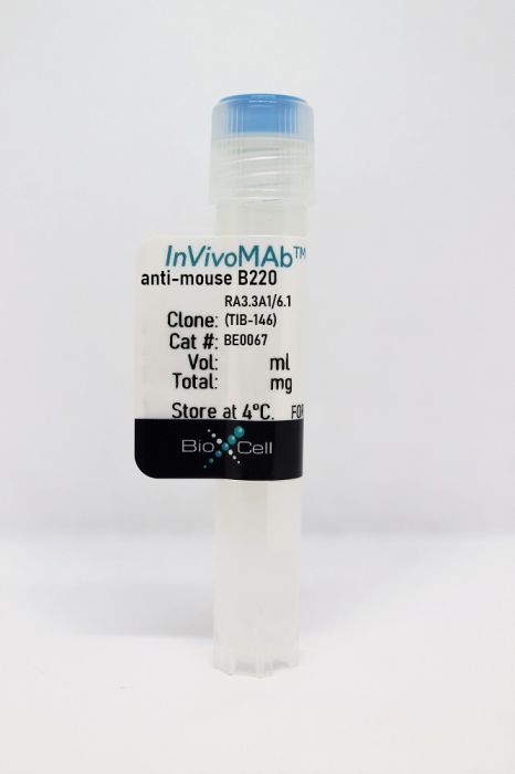
InVivoMAb anti-mouse B220
Product Details
The RA3.3A1/6.1 monoclonal antibody reacts with mouse B220 also known as CD45R. B220 is a 220 kDa transmembrane protein tyrosine phosphatase expressed on B cells and some subsets of T and NK cells. B220 plays a critical role in TCR and BCR signaling and is commonly used as a B cell marker. The RA3.3A1/6.1 antibody is commonly used for in vivo B cell depletion.Specifications
| Isotype | Rat IgM |
|---|---|
| Recommended Isotype Control(s) | InVivoMAb polyclonal rat IgG |
| Recommended Dilution Buffer | InVivoPure pH 7.0 Dilution Buffer |
| Conjugation | This product is unconjugated. Conjugation is available via our Antibody Conjugation Services. |
| Immunogen | Mouse RAW 112 lymphosarcoma cells |
| Reported Applications |
in vivo B cell depletion in vitro B cell negative selection |
| Formulation |
PBS, pH 7.0 Contains no stabilizers or preservatives |
| Endotoxin |
<2EU/mg (<0.002EU/μg) Determined by LAL gel clotting assay |
| Purity |
>95% Determined by SDS-PAGE |
| Sterility | 0.2 µm filtration |
| Production | Purified from cell culture supernatant in an animal-free facility |
| Purification | Protein A |
| RRID | AB_1107651 |
| Storage | The antibody solution should be stored at the stock concentration at 4°C. Do not freeze. |
Recommended Products
-
Recommended Isotype Control(s)
InVivoMAb polyclonal rat IgG
-
Recommended Dilution Buffer
InVivoPure pH 7.0 Dilution Buffer
in vivo B cell depletion
Carmi, Y., et al. (2015). "Allogeneic IgG combined with dendritic cell stimuli induce antitumour T-cell immunity" Nature 521(7550): 99-104. PubMed
Whereas cancers grow within host tissues and evade host immunity through immune-editing and immunosuppression, tumours are rarely transmissible between individuals. Much like transplanted allogeneic organs, allogeneic tumours are reliably rejected by host T cells, even when the tumour and host share the same major histocompatibility complex alleles, the most potent determinants of transplant rejection. How such tumour-eradicating immunity is initiated remains unknown, although elucidating this process could provide the basis for inducing similar responses against naturally arising tumours. Here we find that allogeneic tumour rejection is initiated in mice by naturally occurring tumour-binding IgG antibodies, which enable dendritic cells (DCs) to internalize tumour antigens and subsequently activate tumour-reactive T cells. We exploited this mechanism to treat autologous and autochthonous tumours successfully. Either systemic administration of DCs loaded with allogeneic-IgG-coated tumour cells or intratumoral injection of allogeneic IgG in combination with DC stimuli induced potent T-cell-mediated antitumour immune responses, resulting in tumour eradication in mouse models of melanoma, pancreas, lung and breast cancer. Moreover, this strategy led to eradication of distant tumours and metastases, as well as the injected primary tumours. To assess the clinical relevance of these findings, we studied antibodies and cells from patients with lung cancer. T cells from these patients responded vigorously to autologous tumour antigens after culture with allogeneic-IgG-loaded DCs, recapitulating our findings in mice. These results reveal that tumour-binding allogeneic IgG can induce powerful antitumour immunity that can be exploited for cancer immunotherapy.
in vitro B cell negative selection
Bouffi, C., et al. (2015). "Transcription Factor Repertoire of Homeostatic Eosinophilopoiesis" J Immunol 195(6): 2683-2695. PubMed
The production of mature eosinophils (Eos) is a tightly orchestrated process with the aim to sustain normal Eos levels in tissues while also maintaining low numbers of these complex and sensitive cells in the blood. To identify regulators of homeostatic eosinophilopoiesis in mice, we took a global approach to identify genome-wide transcriptome and epigenome changes that occur during homeostasis at critical developmental stages, including Eos-lineage commitment and lineage maturation. Our analyses revealed a markedly greater number of transcriptome alterations associated with Eos maturation (1199 genes) than with Eos-lineage commitment (490 genes), highlighting the greater transcriptional investment necessary for differentiation. Eos-lineage-committed progenitors (EoPs) were noted to express high levels of granule proteins and contain granules with an ultrastructure distinct from that of mature resting Eos. Our analyses also delineated a 976-gene Eos-lineage transcriptome that included a repertoire of 56 transcription factors, many of which have never previously been associated with Eos. EoPs and Eos, but not granulocyte-monocyte progenitors or neutrophils, expressed Helios and Aiolos, members of the Ikaros family of transcription factors, which regulate gene expression via modulation of chromatin structure and DNA accessibility. Epigenetic studies revealed a distinct distribution of active chromatin marks between genes induced with lineage commitment and genes induced with cell maturation during Eos development. In addition, Aiolos and Helios binding sites were significantly enriched in genes expressed by EoPs and Eos with active chromatin, highlighting a potential novel role for Helios and Aiolos in regulating gene expression during Eos development.
Becher, B., et al. (2014). "High-dimensional analysis of the murine myeloid cell system" Nat Immunol 15(12): 1181-1189. PubMed
Advances in cell-fate mapping have revealed the complexity in phenotype, ontogeny and tissue distribution of the mammalian myeloid system. To capture this phenotypic diversity, we developed a 38-antibody panel for mass cytometry and used dimensionality reduction with machine learning-aided cluster analysis to build a composite of murine (mouse) myeloid cells in the steady state across lymphoid and nonlymphoid tissues. In addition to identifying all previously described myeloid populations, higher-order analysis allowed objective delineation of otherwise ambiguous subsets, including monocyte-macrophage intermediates and an array of granulocyte variants. Using mice that cannot sense granulocyte macrophage-colony stimulating factor GM-CSF (Csf2rb(-/-)), which have discrete alterations in myeloid development, we confirmed differences in barrier tissue dendritic cells, lung macrophages and eosinophils. The methodology further identified variations in the monocyte and innate lymphoid cell compartment that were unexpected, which confirmed that this approach is a powerful tool for unambiguous and unbiased characterization of the myeloid system.
in vitro B cell negative selection
Smyth, L. A., et al. (2013). "Tolerogenic Donor-Derived Dendritic Cells Risk Sensitization In Vivo owing to Processing and Presentation by Recipient APCs" J Immunol 190(9): 4848-4860. PubMed
Modification of allogeneic dendritic cells (DCs) through drug treatment results in DCs with in vitro hallmarks of tolerogenicity. Despite these observations, using murine MHC-mismatched skin and heart transplant models, donor-derived drug-modified DCs not only failed to induce tolerance but also accelerated graft rejection. The latter was inhibited by injecting the recipient with anti-CD8 Ab, which removed both CD8(+) T cells and CD8(+) DCs. The discrepancy between in vitro and in vivo data could be explained, partly, by the presentation of drug-modified donor DC MHC alloantigens by recipient APCs and activation of recipient T cells with indirect allospecificity, leading to the induction of alloantibodies. Furthermore, allogeneic MHC molecules expressed by drug-treated DCs were rapidly processed and presented in peptide form by recipient APCs in vivo within hours of DC injection. Using TCR-transgenic T cells, Ag presentation of injected OVA-pulsed DCs was detectable for = 3 d, whereas indirect presentation of MHC alloantigen by recipient APCs led to activation of T cells within 14 h and was partially inhibited by reducing the numbers of CD8(+) DCs in vivo. In support of this observation when mice lacking CD8(+) DCs were pretreated with drug-modified DCs prior to transplantation, skin graft rejection kinetics were similar to those in non-DC-treated controls. Of interest, when the same mice were treated with anti-CD40L blockade plus drug-modified DCs, skin graft survival was prolonged, suggesting endogenous DCs were responsible for T cell priming. Altogether, these findings highlight the risks and limitations of negative vaccination using alloantigen-bearing “tolerogenic” DCs.
in vitro B cell negative selection
Alam, S. and A. J. Sant. (2011). "Infection with seasonal influenza virus elicits CD4 T cells specific for genetically conserved epitopes that can be rapidly mobilized for protective immunity to pandemic H1N1 influenza virus" J Virol 85(24): 13310-13321. PubMed
In recent years, influenza viruses with pandemic potential have been a major concern worldwide. One unresolved issue is how infection or vaccination with seasonal influenza virus strains influences the ability to mount a protective immune response to novel pandemic strains. In this study, we developed a mouse model of primary and secondary influenza infection by using a widely circulating seasonal H1N1 virus and the pandemic strain of H1N1 that emerged in Mexico in 2009, and we evaluated several key issues. First, using overlapping peptide libraries encompassing the entire translated sequences of 5 major influenza virus proteins, we assessed the specificity of CD4 T cell reactivity toward epitopes conserved among H1N1 viruses or unique to the seasonal or pandemic strain by enzyme-linked immunospot (ELISpot) assays. Our data show that CD4 T cells reactive to both virus-specific and genetically conserved epitopes are elicited, allowing separate tracking of these responses. Populations of cross-reactive CD4 T cells generated from seasonal influenza infection were found to expand earlier after secondary infection with the pandemic H1N1 virus than CD4 T cell populations specific for new epitopes. Coincident with this rapid CD4 T cell response was a potentiated neutralizing-antibody response to the pandemic strain and protection from the pathological effects of infection with the pandemic virus. This protection was not dependent on CD8 T cells. Together, our results indicate that exposure to seasonal vaccines and infection elicits CD4 T cells that promote the ability of the mammalian host to mount a protective immune response to pandemic strains of influenza virus.
in vivo B cell depletion
Olkhanud, P. B., et al. (2011). "Tumor-evoked regulatory B cells promote breast cancer metastasis by converting resting CD4(+) T cells to T-regulatory cells" Cancer Res 71(10): 3505-3515. PubMed
Pulmonary metastasis of breast cancer requires recruitment and expansion of T-regulatory cells (Treg) that promote escape from host protective immune cells. However, it remains unclear precisely how tumors recruit Tregs to support metastatic growth. Here we report the mechanistic involvement of a unique and previously undescribed subset of regulatory B cells. These cells, designated tumor-evoked Bregs (tBreg), phenotypically resemble activated but poorly proliferative mature B2 cells (CD19(+) CD25(High) CD69(High)) that express constitutively active Stat3 and B7-H1(High) CD81(High) CD86(High) CD62L(Low) IgM(Int). Our studies with the mouse 4T1 model of breast cancer indicate that the primary role of tBregs in lung metastases is to induce TGF-beta-dependent conversion of FoxP3(+) Tregs from resting CD4(+) T cells. In the absence of tBregs, 4T1 tumors cannot metastasize into the lungs efficiently due to poor Treg conversion. Our findings have important clinical implications, as they suggest that tBregs must be controlled to interrupt the initiation of a key cancer-induced immunosuppressive event that is critical to support cancer metastasis.
- Immunology and Microbiology,
IL-34 empowers regulatory T cells with novel non-canonical function to safeguard brain barrier integrity during neuro-inflammation
Preprint on BioRxiv : the Preprint Server for Biology on 13 September 2024 by Van Hoecke, L., Verreycken, J., et al.
In efforts to find reparative strategies for brain damage, brain-associated regulatory T cells (Tregs) have gained increasing attention in recent years. Beyond their textbook immunoregulatory function, Tregs have emerged as key players in the response to brain trauma and the restoration of damaged brain tissue. Here, we are the first to describe a novel, non-canonical function of Tregs in maintaining the sealing capacity of both the blood-brain barrier (BBB) and the blood-cerebrospinal fluid (CSF) barrier. Moreover, we identified the cytokine IL-34 as a critical determinant in this newly unveiled Treg function. Mechanistically, IL-34 exerts its influence by modulating the expression and localization of the tight junction protein ZO-1 in both BBB endothelial cells and choroid plexus epithelial cells, thereby reinforcing the strength of the brain barriers. Given the well-established notion of leaky brain barriers and the involvement of immunological components in neurological diseases such as Alzheimer’s disease (AD) and multiple sclerosis (MS), we further demonstrate diminished IL-34 expression in Tregs derived from patients with relapsing-remitting MS (RR-MS) and patients with AD and even mild cognitive impairment (MCI). Remarkably, our study reveals the potential of IL-34 treatment in reinstating the integrity of brain barriers within murine models mimicking these neurological disorders. These ground-breaking findings shed light on the intricate relationship between Tregs, IL-34, and the integrity of brain barriers. They offer novel avenues for therapeutic approaches to ameliorate brain barrier dysfunction in the context of neurological disorders.
- COVID-19,
- Immunology and Microbiology
Inhalable SARS-CoV-2 vaccine for single-dose dry powder aerosol immunization
Preprint on Research Square on 8 September 2023 by Ye, T., Jiao, Z., et al.
The coronavirus disease pandemic has fostered major advances in vaccination technologies; however, there are urgent needs for vaccines that induce mucosal immune responses and single-dose, noninvasive administration. Here, we develop an inhalable, single-dose, dry powder aerosol SARS-CoV-2 vaccine that induces potent systemic and mucosal immune responses. Our vaccine encapsulates proteinaceous cholera toxin B subunit-assembled nanoparticles displaying the SARS-CoV-2 RBD antigen within microcapsules of optimal aerodynamic size, and this unique nano-micro coupled structure supports efficient alveoli delivery, sustained antigen release, and antigen-presenting cell uptake, which are favourable features for induction of immune responses. Moreover, our vaccine successfully induces strong production of IgG and IgA, as well as a local T-cell response, collectively conferring effective protection against SARS-CoV-2 in mice, hamsters, and nonhuman primates. Finally, we also demonstrate a “mosaic iteration” of our vaccine that codisplays ancestral and Omicron antigens, extending the breadth of antibody response against cocirculating strains and transmission of the Omicron variant. These findings support our inhalable vaccine as a promising multivalent platform for fighting COVID-19 or other respiratory infectious diseases.
- Mus musculus (House mouse)
CCR4 and CCR7 differentially regulate thymocyte localization with distinct outcomes for central tolerance.
In eLife on 2 June 2023 by Li, Y., Guaman Tipan, P., et al.
PubMed
Central tolerance ensures autoreactive T cells are eliminated or diverted to the regulatory T cell lineage, thus preventing autoimmunity. To undergo central tolerance, thymocytes must enter the medulla to test their T-cell receptors (TCRs) for autoreactivity against the diverse self-antigens displayed by antigen-presenting cells (APCs). While CCR7 is known to promote thymocyte medullary entry and negative selection, our previous studies implicate CCR4 in these processes, raising the question of whether CCR4 and CCR7 play distinct or redundant roles in central tolerance. Here, synchronized positive selection assays, two-photon time-lapse microscopy, and quantification of TCR-signaled apoptotic thymocytes, demonstrate that CCR4 and CCR7 promote medullary accumulation and central tolerance of distinct post-positive selection thymocyte subsets in mice. CCR4 is upregulated within hours of positive selection signaling and promotes medullary entry and clonal deletion of immature post-positive selection thymocytes. In contrast, CCR7 is expressed several days later and is required for medullary localization and negative selection of mature thymocytes. In addition, CCR4 and CCR7 differentially enforce self-tolerance, with CCR4 enforcing tolerance to self-antigens presented by activated APCs, which express CCR4 ligands. Our findings show that CCR7 expression is not synonymous with medullary localization and support a revised model of central tolerance in which CCR4 and CCR7 promote early and late stages of negative selection, respectively, via interactions with distinct APC subsets. © 2023, Li, Guaman Tipan et al.
- Cancer Research,
- Immunology and Microbiology
Anti-4-1BB immunotherapy enhances systemic immune effects of radiotherapy to induce B and T cell-dependent anti-tumor immune activation and improve tumor control at unirradiated sites.
In Cancer Immunology, Immunotherapy : CII on 1 June 2023 by Martin, A. L., Powell, C., et al.
Radiation therapy (RT) can prime and boost systemic anti-tumor effects via STING activation, resulting in enhanced tumor antigen presentation and antigen recognition by T cells. It is increasingly recognized that optimal anti-tumor immune responses benefit from coordinated cellular (T cell) and humoral (B cell) responses. However, the nature and functional relevance of the RT-induced immune response are controversial, beyond STING signaling, and agonistic interventions are lacking. Here, we show that B and CD4+ T cell accumulation at tumor beds in response to RT precedes the arrival of CD8+ T cells, and both cell types are absolutely required for abrogated tumor growth in non-irradiated tumors. Further, RT induces increased expression of 4-1BB (CD137) in both T and B cells; both in preclinical models and in a cohort of patients with small cell lung cancer treated with thoracic RT. Accordingly, the combination of RT and anti-41BB therapy leads to increased immune cell infiltration in the tumor microenvironment and significant abscopal effects. Thus, 4-1BB therapy enhances radiation-induced tumor-specific immune responses via coordinated B and T cell responses, thereby preventing malignant progression at unirradiated tumor sites. These findings provide a rationale for combining RT and 4-1bb therapy in future clinical trials. © 2022. The Author(s), under exclusive licence to Springer-Verlag GmbH Germany, part of Springer Nature.
- Genetics,
- Immunology and Microbiology
Immune profiling of adeno-associated virus response identifies B cell-specific targets that enable vector re-administration in mice.
In Gene Therapy on 1 May 2023 by Chen, M., Kim, B., et al.
PubMed
Adeno-associated virus (AAV) vector-based gene therapies can be applied to a wide range of diseases. AAV expression can last for months to years, but vector re-administration may be necessary to achieve life-long treatment. Unfortunately, immune responses against these vectors are potentiated after the first administration, preventing the clinical use of repeated administration of AAVs. Reducing the immune response against AAVs while minimizing broad immunosuppression would improve gene delivery efficiency and long-term safety. In this study, we quantified the contributions of multiple immune system components of the anti-AAV response in mice. We identified B-cell-mediated immunity as a critical component preventing vector re-administration. Additionally, we found that IgG depletion alone was insufficient to enable re-administration, suggesting IgM antibodies play an important role in the immune response against AAV. Further, we found that AAV-mediated transduction is improved in µMT mice that lack functional IgM heavy chains and cannot form mature B-cells relative to wild-type mice. Combined, our results suggest that B-cells, including non-class switched B-cells, are a potential target for therapeutics enabling AAV re-administration. Our results also suggest that the µMT mice are a potentially useful experimental model for gene delivery studies since they allow repeated dosing for more efficient gene delivery from AAVs. © 2022. The Author(s), under exclusive licence to Springer Nature Limited.
- Immunology and Microbiology
GPR55 in B cells limits atherosclerosis development and regulates plasma cell maturation.
In Nat Cardiovasc Res on 1 November 2022 by Guillamat-Prats, R., Hering, D., et al.
PubMed
Dissecting the pathways regulating the adaptive immune response in atherosclerosis is of particular therapeutic interest. Here we report that the lipid G-protein coupled receptor GPR55 is highly expressed by splenic plasma cells (PC), upregulated in mouse spleens during atherogenesis and human unstable or ruptured compared to stable plaques. Gpr55-deficient mice developed larger atherosclerotic plaques with increased necrotic core size compared to their corresponding controls. Lack of GPR55 hyperactivated B cells, disturbed PC maturation and resulted in immunoglobulin (Ig)G overproduction. B cell-specific Gpr55 depletion or adoptive transfer of Gpr55-deficient B cells was sufficient to promote plaque development and elevated IgG titers. In vitro, the endogenous GPR55 ligand lysophsophatidylinositol (LPI) enhanced PC proliferation, whereas GPR55 antagonism blocked PC maturation and increased their mitochondrial content. Collectively, these discoveries provide previously undefined evidence for GPR55 in B cells as a key modulator of the adaptive immune response in atherosclerosis.
Targeted immunosuppression enhances repeated gene delivery
Preprint on Research Square on 1 March 2022 by Chen, M., Kim, B., et al.
PubMed
Adeno-associated virus (AAV) vector-based gene therapies can be applied to a wide range of diseases. AAV expression can last for months to years, but vector re-administration may be necessary to achieve life-long treatment. Unfortunately, immune system response against these vectors is potentiated after the first administration, which prevents the clinical use of repeated administration of AAVs. Reducing immune response against AAVs while minimizing immunosuppression would improve gene delivery efficiency and long-term safety. In this study, we quantified the contributions of multiple immune system components towards AAV response in mice. We identified B-cell-mediated immunity as a critical component preventing vector re-administration. Specifically, we found that IgG depletion was insufficient to enhance re-administration, suggesting the key role of B-cell mediated IgM antibodies in the immune response against AAV. Further, we also found that AAV-mediated transduction is improved compared to wild-type mice in µMT mice that lack functional IgM heavy chains and cannot form mature B-cells. Combined, our results suggest that IgM production in B cells is a potential target for therapeutics enabling AAV re-administration. Our results also suggest that the µMT mice are a potentially useful experimental model for gene delivery studies since they allow for up to 15-fold more efficient gene delivery.
- Immunology and Microbiology
GPR55 in B cells limits atherosclerosis development and regulates plasma cell maturation
Preprint on Research Square on 12 January 2022 by Guillamat-Prats, R., Hering, D., et al.
PubMed
Identifying novel pathways regulating the adaptive immune response in chronic inflammatory diseases such as atherosclerosis is of particular interest in view of developing new therapeutic drugs. Here we report that the lipid receptor GPR55 is highly expressed by splenic B cells and inversely correlates with atheroma plaque size in mice. In human carotid endarterectomy specimen, GPR55 transcript levels were significantly lower in unstable compared to stable carotid plaques. To study the impact of GPR55 deficiency in atherosclerosis, we crossed Gpr55 knockout mice with apolipoprotein E (ApoE) knockout mice and subjected the mice to Western diet for 4 to 16 weeks. Compared to ApoE-/- controls, ApoE-/-Gpr55-/- mice developed larger plaques with increased necrotic core size, associated with elevated circulating and aortic leukocyte counts. Flow cytometry, immunofluorescence and RNA-sequencing analysis of splenic B cells in these mice revealed a hyperactivated B cell phenotype with disturbed plasma cell maturation and immunoglobulin (Ig)G antibody overproduction. The specific contribution of B cell GPR55 in atherosclerosis was further studied in mixed Gpr55-/-/µMT bone marrow chimeras on low density receptor deficiency (Ldlr-/-) background, revealing that B-cell specific depletion of Gpr55 was sufficient to promote plaque development. Conversely, adoptive transfer of wildtype B cells into ApoE-/-Gpr55-/- mice blunted the proatherogenic phenotype. In vitro stimulation of splenocytes with the endogenous GPR55 ligand LPI promoted plasma cell proliferation and enhanced B cell activation marker expression, which was inhibited by the GPR55 antagonist CID16020046. Collectively, these discoveries provide new evidence for GPR55 as key modulator of the adaptive immune response in atherosclerosis. Targeting GPR55 could be useful to limit inflammation and plaque progression in patients suffering from atherosclerosis.
- IP,
- Mus musculus (House mouse),
- Immunology and Microbiology
TGF-β-mediated silencing of genomic organizer SATB1 promotes Tfh cell differentiation and formation of intra-tumoral tertiary lymphoid structures.
In Immunity on 11 January 2022 by Chaurio, R. A., Anadon, C. M., et al.
PubMed
The immune checkpoint receptor PD-1 on T follicular helper (Tfh) cells promotes Tfh:B cell interactions and appropriate positioning within tissues. Here, we examined the impact of regulation of PD-1 expression by the genomic organizer SATB1 on Tfh cell differentiation. Vaccination of CD4CreSatb1f/f mice enriched for antigen-specific Tfh cells, and TGF-β-mediated repression of SATB1 enhanced Tfh differentiation of human T cells. Mechanistically, high Icos expression in Satb1-/- CD4+ T cells promoted Tfh cell differentiation by preventing T follicular regulatory cell skewing and resulted in increased isotype-switched B cell responses in vivo. Ovarian tumors in CD4CreSatb1f/f mice accumulated tumor antigen-specific, LIGHT+CXCL13+IL-21+ Tfh cells and tertiary lymphoid structures (TLS). TLS formation decreased tumor growth in a CD4+ T cell and CXCL13-dependent manner. The transfer of Tfh cells, but not naive CD4+ T cells, induced TLS at tumor beds and decreased tumor growth. Thus, TGF-β-mediated silencing of Satb1 licenses Tfh cell differentiation, providing insight into the genesis of TLS within tumors. Copyright © 2021 The Author(s). Published by Elsevier Inc. All rights reserved.
- Immunology and Microbiology
GPR55 in B cells limits atherosclerosis development and regulates plasma cell maturation
Preprint on BioRxiv : the Preprint Server for Biology on 21 December 2021 by Guillamat-Prats, R., Hering, D., et al.
PubMed
Identifying novel pathways regulating the adaptive immune response in chronic inflammatory diseases such as atherosclerosis is of particular interest in view of developing new therapeutic drugs. Here we report that the lipid receptor GPR55 is highly expressed by splenic B cells and inversely correlates with atheroma plaque size in mice. In human carotid endarterectomy specimen, GPR55 transcript levels were significantly lower in unstable compared to stable carotid plaques. To study the impact of GPR55 deficiency in atherosclerosis, we crossed Gpr55 knockout mice with apolipoprotein E ( ApoE ) knockout mice and subjected the mice to Western diet for 4 to 16 weeks. Compared to ApoE -/- controls, ApoE -/- Gpr55 -/- mice developed larger plaques with increased necrotic core size, associated with elevated circulating and aortic leukocyte counts. Flow cytometry, immunofluorescence and RNA-sequencing analysis of splenic B cells in these mice revealed a hyperactivated B cell phenotype with disturbed plasma cell maturation and immunoglobulin (Ig)G antibody overproduction. The specific contribution of B cell GPR55 in atherosclerosis was further studied in mixed Gpr55 -/- / µMT bone marrow chimeras on low density receptor deficiency ( Ldlr -/- ) background, revealing that B-cell specific depletion of Gpr55 was sufficient to promote plaque development. Conversely, adoptive transfer of wildtype B cells into ApoE -/- Gpr55 -/- mice blunted the proatherogenic phenotype. In vitro stimulation of splenocytes with the endogenous GPR55 ligand LPI promoted plasma cell proliferation and enhanced B cell activation marker expression, which was inhibited by the GPR55 antagonist CID16020046. Collectively, these discoveries provide new evidence for GPR55 as key modulator of the adaptive immune response in atherosclerosis. Targeting GPR55 could be useful to limit inflammation and plaque progression in patients suffering from atherosclerosis.
- Biochemistry and Molecular biology,
- Genetics,
- Immunology and Microbiology,
- Neuroscience
Single-cell RNA sequencing reveals B cell-related molecular biomarkers for Alzheimer's disease.
In Experimental & Molecular Medicine on 1 December 2021 by Xiong, L. L., Xue, L. L., et al.
PubMed
In recent years, biomarkers have been integrated into the diagnostic process and have become increasingly indispensable for obtaining knowledge of the neurodegenerative processes in Alzheimer's disease (AD). Peripheral blood mononuclear cells (PBMCs) in human blood have been reported to participate in a variety of neurodegenerative activities. Here, a single-cell RNA sequencing analysis of PBMCs from 4 AD patients (2 in the early stage, 2 in the late stage) and 2 normal controls was performed to explore the differential cell subpopulations in PBMCs of AD patients. A significant decrease in B cells was detected in the blood of AD patients. Furthermore, we further examined PBMCs from 43 AD patients and 41 normal subjects by fluorescence activated cell sorting (FACS), and combined with correlation analysis, we found that the reduction in B cells was closely correlated with the patients' Clinical Dementia Rating (CDR) scores. To confirm the role of B cells in AD progression, functional experiments were performed in early-stage AD mice in which fibrous plaques were beginning to appear; the results demonstrated that B cell depletion in the early stage of AD markedly accelerated and aggravated cognitive dysfunction and augmented the Aβ burden in AD mice. Importantly, the experiments revealed 18 genes that were specifically upregulated and 7 genes that were specifically downregulated in B cells as the disease progressed, and several of these genes exhibited close correlation with AD. These findings identified possible B cell-based AD severity, which are anticipated to be conducive to the clinical identification of AD progression. © 2021. The Author(s).
- Cancer Research,
- Immunology and Microbiology
Redefining innate natural antibodies as important contributors to anti-tumor immunity.
In eLife on 5 October 2021 by Rawat, K., Tewari, A., et al.
PubMed
Myeloid, T, and NK cells are key players in the elimination phase of cancer immunoediting, also referred to as cancer immunosurveillance. However, the role of B cells and NAbs, which are present prior to the encounter with cognate antigens, has been overlooked. One reason is due to the popular use of a single B cell-deficient mouse model, muMT mice. Cancer models using muMT mice display a similar tumor burden as their wild-type (WT) counterparts. Empirically, we observe what others have previously reported with muMT mice. However, using two other B cell-deficient mouse models (IgHELMD4 and CD19creDTA), we show a three- to fivefold increase in tumor burden relative to WT mice. In addition, using an unconventional, non-cancer-related, immune neoantigen model where hypoxic conditions and cell clustering are absent, we provide evidence that B cells and their innate, natural antibodies (NAbs) are critical for the detection and elimination of neoantigen-expressing cells. Finally, we find that muMT mice display anti-tumor immunity because of an unexpected compensatory mechanism consisting of significantly enhanced type 1 interferon (IFN)-producing plasmacytoid dendritic cells (pDCs), which recruit a substantial number of NK cells to the tumor microenvironment compared to WT mice. Diminishing this compensatory pDC-IFN-NK cell mechanism revealed that muMT mice develop a three- to fivefold increase in tumor burden compared to WT mice. In summary, our findings suggest that NAbs are part of an early defense against not only microorganisms and dying cells, but precancerous cells as well. © 2021, Rawat et al.
- Cancer Research,
- Immunology and Microbiology
Targeted Deletion of CXCR2 in Myeloid Cells Alters the Tumor Immune Environment to Improve Antitumor Immunity.
In Cancer Immunology Research on 1 February 2021 by Yang, J., Yan, C., et al.
PubMed
Recruitment of myeloid-derived suppressor cells (MDSC) into the tumor microenvironment (TME) contributes to cancer immune evasion. MDSCs express the chemokine receptor CXCR2, and inhibiting CXCR2 suppresses the recruitment of MDSCs into the tumor and the premetastatic niche. Here, we compared the growth and metastasis of melanoma and breast cancer xenografts in mice exhibiting or not exhibiting targeted deletion of Cxcr2 in myeloid cells (CXCR2myeΔ/Δ vs. CXCR2myeWT). Detailed analysis of leukocyte populations in peripheral blood and in tumors from CXCR2myeΔ/Δ mice revealed that loss of CXCR2 signaling in myeloid cells resulted in reduced intratumoral MDSCs and increased intratumoral CXCL11. The increase in intratumoral CXCL11 was derived in part from tumor-infiltrating B1b cells. The reduction in intratumoral MDSCs coupled with an increase in intratumoral B1b cells expressing CXCL11 resulted in enhanced infiltration and activation of effector CD8+ T cells in the TME of CXCR2myeΔ/Δ mice, accompanied by inhibition of tumor growth in CXCR2myeΔ/Δ mice compared with CXCR2myeWT littermates. Treatment of tumor-bearing mice with a CXCR2 antagonist (SX-682) also inhibited tumor growth, reduced intratumoral MDSCs, and increased intratumoral B1b cells expressing CXCL11, leading to an increase in activated CD8+ T cells in the tumor. Depletion of B220+ cells or depletion of CD8+ T cells reversed the tumor-inhibitory properties in CXCR2myeΔ/Δ mice. These data revealed a mechanism by which loss of CXCR2 signaling in myeloid cells modulates antitumor immunity through decreasing MDSCs and enriching CXCL11-producing B1b cells in the TME, which in turn increases CD8+ T-cell recruitment and activation in tumors. ©2020 American Association for Cancer Research.
- In Vivo,
- Mus musculus (House mouse),
- Immunology and Microbiology,
- Neuroscience
In situ Vaccine Plus Checkpoint Blockade Induces Memory Humoral Response.
In Frontiers in Immunology on 28 August 2020 by Baniel, C. C., Heinze, C. M., et al.
PubMed
In a syngeneic murine melanoma (MEL) model, we recently reported an in situ vaccination response to combined radiation (RT) and intra-tumoral (IT) injection of anti-GD2 hu14. 18-IL2 immunocytokine (IC). This combined treatment resulted in 71% complete and durable regression of 5-week tumors, a tumor-specific memory T cell response, and augmented response to systemic anti-CTLA-4 antibody checkpoint blockade. While the ability of radiation to diversify anti-tumor T cell response has been reported, we hypothesize that mice rendered disease-free (DF) by a RT-based ISV might also exhibit a heightened B cell response. C57BL/6 mice were engrafted with 2 × 10⁶ GD2+ B78 MEL and treated at a target tumor size of ~200 mm3 with 12 Gy RT, IT-IC on day (D)6-D10, and anti-CTLA-4 on D3, 6, and 9. Serum was collected via facial vein before tumor injection, before treatment, during treatment, after becoming DF, and following rejection of subcutaneous 2 × 10⁶ B78 MEL re-challenge on D90. Flow cytometry demonstrated the presence of tumor-specific IgG in sera from mice rendered DF and rejecting re-challenge with B78 MEL at D90 after starting treatment. Consistent with an adaptive endogenous anti-tumor humoral memory response, these anti-tumor antibodies bound to B78 cells and parental B16 cells (GD2-), but not to the unrelated syngeneic Panc02 or Panc02 GD2+ cell lines. We evaluated the kinetics of this response and observed that tumor-specific IgG was consistently detected by D22 after initiation of treatment, corresponding to a time of rapid tumor regression. The amount of tumor-specific antibody binding to tumor cells (as measured by flow MFI) did not correlate with host animal prognosis. Incubation of B16 MEL cells in DF serum, vs. naïve serum, prior to IV injection, did not delay engraftment of B16 metastases and showed similar overall survival rates. B cell depletion using anti-CD20 or anti-CD19 and anti-B220 did not impact the efficacy of ISV treatment. Thus, treatment with RT + IC + anti-CTLA-4 results in adaptive anti-tumor humoral memory response. This endogenous tumor-specific antibody response does not appear to have therapeutic efficacy but may serve as a biomarker for an anti-tumor T cell response. Copyright © 2020 Baniel, Heinze, Hoefges, Sumiec, Hank, Carlson, Jin, Patel, Sriramaneni, Gillies, Erbe, Schwarz, Pieper, Rakhmilevich, Sondel and Morris.
- In Vivo,
- Mus musculus (House mouse),
- Immunology and Microbiology
Complement Signals Determine Opposite Effects of B Cells in Chemotherapy-Induced Immunity.
In Cell on 19 March 2020 by Lu, Y., Zhao, Q., et al.
PubMed
Understanding molecular mechanisms that dictate B cell diversity is important for targeting B cells as anti-cancer treatment. Through the single-cell dissection of B cell heterogeneity in longitudinal samples of patients with breast cancer before and after neoadjuvant chemotherapy, we revealed that an ICOSL+ B cell subset emerges after chemotherapy. Using three immunocompetent mouse models, we recapitulated the subset switch of human tumor-infiltrating B cells during chemotherapy. By employing B-cell-specific deletion mice, we showed that ICOSL in B cells boosts anti-tumor immunity by enhancing the effector to regulatory T cell ratio. The signature of ICOSL+ B cells is imprinted by complement-CR2 signaling, which is triggered by immunogenic cell death. Moreover, we identified that CD55, a complement inhibitory protein, determines the opposite roles of B cells in chemotherapy. Collectively, we demonstrated a critical role of the B cell subset switch in chemotherapy response, which has implications in designing novel anti-cancer therapies. VIDEO ABSTRACT. Copyright © 2020 Elsevier Inc. All rights reserved.
- Immunology and Microbiology
Toxoplasma gondii tkl1 Deletion Mutant Is a Promising Vaccine against Acute, Chronic, and Congenital Toxoplasmosis in Mice.
In The Journal of Immunology on 15 March 2020 by Wang, J. L., Liang, Q. L., et al.
PubMed
In this study, we generated a tkl1 deletion mutant in the Toxoplasma gondii type 1 RH (RHΔtkl1) strain and tested the protective efficacies of vaccination using RHΔtkl1 tachyzoites against acute, chronic, and congenital T. gondii infections in Kunming mice. Mice vaccinated with RHΔtkl1 mounted a strong humoral and cellular response as shown by elevated levels of anti-T. gondii-specific IgG, IL-2, IL-12, IFN-γ, and IL-10. All RHΔtkl1-vaccinated mice survived a lethal challenge with 1 × 10³ tachyzoites of type 1 RH or ToxoDB#9 (PYS or TgC7) strain as well as 100 cysts or oocysts of Prugniuad strain. All mock-vaccinated plus infected mice have died. Vaccination also protected against cyst- or oocyst-caused chronic infection, reduced vertical transmission caused by oocysts, increased litter size, and maintained body weight of pups born to dams challenged with 10 oocysts on day 5 of gestation. In contrast, all mock-vaccinated plus oocysts-infected dams had aborted, and no fetus has survived. Vaccinated dams remained healthy postinfection, and their brain cyst burden was significantly reduced compared with mock-vaccinated dams infected with oocysts. In vivo depletion of CD4+ T cells, CD8+ T cells, and B cells revealed that CD8+ T cells are involved in the protection of mice against T. gondii infection. Additionally, adoptive transfer of CD8+ T cells from RHΔtkl1-vaccinated mice significantly enhanced the survival of naive mice infected with the pathogenic strain. Together, these data reaffirm the importance of CD8+ T cell responses in future vaccine design for toxoplasmosis and present T. gondii tkl1 gene as a promising vaccine candidate. Copyright © 2020 by The American Association of Immunologists, Inc.
- FC/FACS,
- Mus musculus (House mouse),
- Immunology and Microbiology
Live-cell imaging reveals the relative contributions of antigen-presenting cell subsets to thymic central tolerance.
In Nature Communications on 17 May 2019 by Lancaster, J. N., Thyagarajan, H. M., et al.
PubMed
Both medullary thymic epithelial cells (mTEC) and dendritic cells (DC) present tissue-restricted antigens (TRA) to thymocytes to induce central tolerance, but the relative contributions of these antigen-presenting cell (APC) subsets remain unresolved. Here we developed a two-photon microscopy approach to observe thymocytes interacting with intact APCs presenting TRAs. We find that mTECs and DCs cooperate extensively to induce tolerance, with their relative contributions regulated by the cellular form of the TRA and the class of major histocompatibility complex (MHC) on which antigen is presented. Even when TRA expression is restricted to mTECs, DCs still present self-antigens at least as frequently as mTECs. Notably, the DC subset cDC2 efficiently acquires secreted mTEC-derived TRAs for cross-presentation on MHC-I. By directly imaging interactions between thymocytes and APCs, while monitoring intracellular signaling, this study reveals that distinct DC subsets and AIRE+ mTECs contribute substantially to presentation of diverse self-antigens for establishing central tolerance.
- Immunology and Microbiology,
- In Vivo,
- Mus musculus (House mouse)
A Subset of Type I Conventional Dendritic Cells Controls Cutaneous Bacterial Infections through VEGFα-Mediated Recruitment of Neutrophils.
In Immunity on 16 April 2019 by Janela, B., Patel, A. A., et al.
PubMed
Skin conventional dendritic cells (cDCs) exist as two distinct subsets, cDC1s and cDC2s, which maintain the balance of immunity to pathogens and tolerance to self and microbiota. Here, we examined the roles of dermal cDC1s and cDC2s during bacterial infection, notably Propionibacterium acnes (P. acnes). cDC1s, but not cDC2s, regulated the magnitude of the immune response to P. acnes in the murine dermis by controlling neutrophil recruitment to the inflamed site and survival and function therein. Single-cell mRNA sequencing revealed that this regulation relied on secretion of the cytokine vascular endothelial growth factor α (VEGF-α) by a minor subset of activated EpCAM+CD59+Ly-6D+ cDC1s. Neutrophil recruitment by dermal cDC1s was also observed during S. aureus, bacillus Calmette-Guérin (BCG), or E. coli infection, as well as in a model of bacterial insult in human skin. Thus, skin cDC1s are essential regulators of the innate response in cutaneous immunity and have roles beyond classical antigen presentation.Copyright © 2019 Elsevier Inc. All rights reserved.
- Cancer Research
IL-33 delays metastatic peritoneal cancer progression inducing an allergic microenvironment.
In Oncoimmunology on 14 December 2018 by Perales-Puchalt, A., Svoronos, N., et al.
PubMed
Ovarian cancer is frequently diagnosed as peritoneal carcinomatosis. Unlike other tumor locations, the peritoneal cavity is commonly exposed to gut-breaching and ascending genital microorganisms and has a unique immune environment. IL-33 is a local cytokine that can activate innate and adaptive immunity. We studied the effectiveness of local IL-33 delivery in the treatment of cancer that has metastasized to the peritoneal cavity. Direct peritoneal administration of IL-33 delayed the progression of metastatic peritoneal cancer. Prolongation in survival was not associated with a direct effect of IL-33 on tumor cells, but with major changes in the immune microenvironment of the tumor. IL-33 promoted a significant increase in the leukocyte compartment of the tumor immunoenvironment and an allergic cytokine profile. We observed a substantial increase in the number of activated CD4+ T-cells accompanied by peritoneal eosinophil infiltration, B-cell activation and activation of peritoneal macrophages which displayed tumoricidal capacity. Depletion of CD4+ cells, eosinophils or macrophages reduced the anti-tumor effects of IL-33 but none of these alone were sufficient to completely abrogate its positive benefit. In conclusion, local administration of IL-33 generates an allergic tumor environment resulting in a novel approach for treatment of metastatic peritoneal malignancies, such as advanced ovarian cancer.
- Cancer Research,
- Immunology and Microbiology
Homoharringtonine induced immune alteration for an Efficient Anti-tumor Response in Mouse Models of Non-small Cell Lung Adenocarcinoma Expressing Kras Mutation.
In Scientific Reports on 29 May 2018 by Weng, T. Y., Wu, H. F., et al.
PubMed
Homoharringtonine (HHT), an inhibitor of protein synthesis, has been used to treat leukemia. Its therapeutic effects on non-small cell lung adenocarcinoma carrying KRAS mutation and their immune system are less understood. The present study examined the therapeutic efficacy and the immune effects of HHT in two murine lung tumor models, xenograft and transgenic, carrying the Kras mutation G12D and G12C respectively. HHT exhibited efficient anticancer activity, significantly suppressing lung tumor growth in vitro and in vivo. The levels of 22 cytokines and chemokines in splenocytes of tumor-bearing mice were examined. Interleukin-12 expression was lower in splenocytes of HHT-treated mice when compared to the controls as demonstrated by a cytokine array and an enzyme-linked immunosorbent assay. The expression levels of CD80, CD86, and CD69 in B220+ B cells from splenocytes of HHT-treated mice were higher than that of control mice in two mouse tumor models. Furthermore, antitumor effect of HHT was attenuated with depletion of B cells. Increased numbers of CD80+ and CD86+ B cells were observed in the mice treated with narciclasine, another translation inhibitor. In conclusion, HHT changed the features of immune cells, and exhibited efficient anti-tumor activity against lung tumor carrying mutant Kras expression.