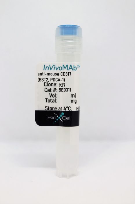
InVivoMAb anti-mouse CD317 (BST2, PDCA-1)
Product Details
The 927 monoclonal antibody reacts with mouse CD317 also known as BST2 and PDCA-1, a 29-33 kDa type II transmembrane glycoprotein. CD317 is expressed exclusively by plasmacytoid dendritic cells and serves as a marker for these cells. CD317 is also sometimes expressed by some tumor cells, including multiple myeloma, renal cell carcinoma, and melanoma cells. The 927 antibody is a useful tool to specifically deplete plasmacytoid dendritic cells when administered in vivo.Specifications
| Isotype | Rat IgG2b, κ |
|---|---|
| Recommended Isotype Control(s) | InVivoMAb rat IgG2b isotype control, anti-keyhole limpet hemocyanin |
| Recommended Dilution Buffer | InVivoPure pH 7.0 Dilution Buffer |
| Conjugation | This product is unconjugated. Conjugation is available via our Antibody Conjugation Services. |
| Immunogen | Mouse type I IFN-producing cells |
| Reported Applications |
in vivo pDC depletion Immunofluorescence Flow cytometry |
| Formulation |
PBS, pH 7.0 Contains no stabilizers or preservatives |
| Endotoxin |
<2EU/mg (<0.002EU/μg) Determined by LAL gel clotting assay |
| Purity |
>95% Determined by SDS-PAGE |
| Sterility | 0.2 µm filtration |
| Production | Purified from cell culture supernatant in an animal-free facility |
| Purification | Protein G |
| RRID | AB_2736991 |
| Molecular Weight | 150 kDa |
| Storage | The antibody solution should be stored at the stock concentration at 4°C. Do not freeze. |
Recommended Products
-
Recommended Isotype Control(s)
InVivoMAb rat IgG2b isotype control, anti-keyhole limpet hemocyanin
-

Recommended Dilution Buffer
InVivoPure pH 7.0 Dilution Buffer
in vivo pDC depletion
Bradley, K. C., et al. (2019). "Microbiota-Driven Tonic Interferon Signals in Lung Stromal Cells Protect from Influenza Virus Infection" Cell Rep 28(1): 245-256.e244. PubMed
Type I interferon (IFNα/β) pathways are fine-tuned to elicit antiviral protection while minimizing immunopathology; however, the initiating stimuli, target tissues, and underlying mechanisms are unclear. Using models of physiological and dysregulated IFNα/β receptor (IFNAR1) surface expression, we show here that IFNAR1-dependent signals set the steady-state IFN signature in both hematopoietic and stromal cells. Increased IFNAR1 levels promote a lung environment refractory to early influenza virus replication by elevating the baseline interferon signature. Commensal microbiota drive the IFN signature specifically in lung stroma, as shown by antibiotic treatment and fecal transplantation. Bone marrow chimera experiments identify lung stromal cells as crucially important for early antiviral immunity and stroma-immune cell interaction for late antiviral resistance. We propose that the microbiota-driven interferon signature in lung epithelia impedes early virus replication and that IFNAR1 surface levels fine-tune this signature. Our findings highlight the interplay between bacterial and viral exposure, with important implications for antibiotic use.
in vivo pDC depletion
Nash, W. T., et al. (2017). "Murine Cytomegalovirus Disrupts Splenic Dendritic Cell Subsets via Type I Interferon-Dependent and -Independent Mechanisms" Front Immunol 8: 251. PubMed
Dendritic cells (DC) are well-known modulators of immunity. This heterogeneous population is composed of defined subsets that exhibit functional specialization and are critical in initiating responses to pathogens. As such, many infectious agents employ strategies to disrupt DC functioning in attempts to evade the immune system. In some instances, this manifests as an outright loss of these cells. Previous work has suggested that, in the absence of an efficient natural killer (NK) cell response, murine cytomegalovirus (MCMV) induces large amounts of interferon (IFN)-I. This heightened IFN-I response is thought to contribute to conventional DC (cDC) loss and delayed development of T cell immunity. However, the precise role of IFN-I in such cDC loss remains unclear. We investigated the effects of licensed NK cells and IFN-I signaling on splenic cDC subsets during MCMV infection and found that a licensed NK cell response partially protects cDC numbers, but does not prevent increases in serum IFN-I. This suggested that high residual IFN-I could contribute to cDC loss. Therefore, we used multiple strategies to modulate IFN-I signaling during MCMV infection including plasmacytoid DC depletion, IFN-I receptor (IFNAR) blockade, and genetic ablation of IFNAR expression. Interestingly, restriction of IFN-I signals did not substantially preserve either CD8(+) or CD4(+) DC total numbers, but resulted in significant retention and/or accumulation of the splenic CD8(-) CD4(-) [double negative (DN)] subset. However, the DN DC effect manifested in a DC-extrinsic manner since IFNAR-deficient cells were not preferentially retained over their IFNAR wild-type counterparts in a mixed-chimera setting. Our results show that IFN-I signaling is not responsible for overt cDC toxicity in the setting of acute MCMV infection and emphasize that additional mechanisms contribute to DC loss and require exploration.
Flow Cytometry
Toivonen, R., et al. (2016). "Activation of Plasmacytoid Dendritic Cells in Colon-Draining Lymph Nodes during Citrobacter rodentium Infection Involves Pathogen-Sensing and Inflammatory Pathways Distinct from Conventional Dendritic Cells" J Immunol 196(11): 4750-4759. PubMed
Dendritic cells (DCs) bear the main responsibility for initiation of adaptive immune responses necessary for antimicrobial immunity. In the small intestine, afferent lymphatics convey Ags and microbial signals to mesenteric lymph nodes (LNs) to induce adaptive immune responses against microbes and food Ags derived from the small intestine. Whether the large intestine is covered by the same lymphatic system or represents its own lymphoid compartment has not been studied until very recently. We identified three small mesenteric LNs, distinct from small intestinal LNs, which drain lymph specifically from the colon, and studied DC responses to the attaching and effacing pathogen Citrobacter rodentium in these. Transcriptional profiling of conventional (CD11c(high)CD103(high)) DC and plasmacytoid (plasmacytoid DC Ag-1(high)B220(+)CD11c(int)) DC (pDC) populations during steady-state conditions revealed activity of distinct sets of genes in these two DC subsets, both in small intestinal and colon-draining LNs. C. rodentium activated DC especially in colon-draining LNs, and gene expression changed in pDC more profoundly than in conventional DC. Among the genes most upregulated in pDC were C-type lectin receptor CLEC4E, IL-1Rs (IL-1R1 and -2), proinflammatory cytokines (IL-1a and IL-6), and TLR6. Our results indicate that colon immune surveillance is distinct from that of the small intestine in terms of draining LNs, and identify pDC as active sentinels of colonic inflammation and/or microbial dysbiosis.
Immunofluorescence
Schliemann, C., et al. (2010). "In vivo biotinylation of the vasculature in B-cell lymphoma identifies BST-2 as a target for antibody-based therapy" Blood 115(3): 736-744. PubMed
The discovery of accessible markers of lymphoma may facilitate the development of antibody-based therapeutic strategies. Here, we describe the results of a chemical proteomic study, based on the in vivo biotinylation of vascular proteins in lymphoma-bearing mice followed by mass spectrometric and bioinformatic analysis, to discover proteins expressed at the tissue-blood border of disseminated B-cell lymphoma. From a list of 58 proteins, which were more than 10-fold up-regulated in nodal and extranodal lymphoma lesions compared with their levels in the corresponding normal host organs, we validated BST-2 as a novel vascular marker of B-cell lymphoma, using immunochemical techniques and in vivo biodistribution studies. Furthermore, targeting BST-2 with 2 independent monoclonal antibodies delayed lymphoma growth in a syngeneic mouse model of the disease. The results of this study delineate a strategy for the treatment of systemic B-cell lymphoma in humans and suggest that anti-BST-2 antibodies may facilitate pharmacodelivery approaches that target the tumor-stroma interface.
in vivo pDC depletion
Rajagopal, D., et al. (2010). "Plasmacytoid dendritic cell-derived type I interferon is crucial for the adjuvant activity of Toll-like receptor 7 agonists" Blood 115(10): 1949-1957. PubMed
There is a high demand for the development of adjuvants that induce cytotoxic T lymphocytes, which are crucial for the elimination of intracellular pathogens and tumor cells. Toll-like receptor (TLR) agonists are prime candidates to fulfill this role because they induce innate immune activation and promote adaptive immune responses. The successful application of the TLR7 agonist R837 for treatment of basal cell carcinoma shows the potential for exploiting this pathway in tumor immunotherapy. Imidazoquinolines like R837 and stimulatory ssRNA oligonucleotides both trigger TLR7-mediated immune activation, but little is known about their comparative ability to promote immunity induction. We investigated differences in innate immune activation and adjuvant activity between the imidazoquinoline R848 and the ssRNA TLR7 agonist polyUs21. In contrast to R848, polyUs21 induced detectable levels of intracellular interferon-alpha (IFN-alpha) in plasmacytoid dendritic cells (PDCs). In immunization studies, only polyUs21 led to robust priming of type 1 T helper cells and cytotoxic T lymphocytes, and it was more efficient in inducing antitumor immunity than R848. Notably, exogenous IFN-alpha augmented the adjuvant activity of R848, whereas depletion of PDC abrogated the adjuvanticity of polyUs21. This study, therefore, identifies sufficient IFN-alpha production by PDC as an important determinant of vaccine efficacy.
in vivo pDC depletion
Moniz, R. J., et al. (2010). "Plasmacytoid dendritic cells modulate nonprotective T-cell responses to genital infection by Chlamydia muridarum" FEMS Immunol Med Microbiol 58(3): 397-404. PubMed
Given their immune-modulating capacity, regulatory T cells (Treg) cells may be important players in the induction of the protective T-cell response (Th1) to genital chlamydial infection. Recent work has demonstrated that plasmacytoid dendritic cells (pDC) respond to genital chlamydial infection, and that pDC may be uniquely positioned for the induction of Treg cells during this infection. Here, we present the first data demonstrating that Treg influx into the draining lymph node and the site of infection during genital chlamydial infection. We found that pDC depletion altered the numbers of Treg and nonprotective inflammatory cells [interferongamma-(IFNgamma)-producing CD8+ T and IFNgamma-producing natural killer T cells] in the spleens of mice genitally infected with Chlamydia muridarum. Furthermore, pDC depletion did not alter Th1 cell numbers, indicating that pDC modulate cells that could inhibit and promote nonprotective inflammation during genital chlamydial infection. Finally, we demonstrate that depletion of pDC results in less severe dilation and collagen deposition in the oviduct following resolution of infection, implicating pDC activity in the formation of sequelae following genital C. muridarum infection.
in vivo pDC depletion, Flow Cytometry
Blasius, A. L., et al. (2006). "Bone marrow stromal cell antigen 2 is a specific marker of type I IFN-producing cells in the naive mouse, but a promiscuous cell surface antigen following IFN stimulation" J Immunol 177(5): 3260-3265. PubMed
Type I IFN-producing cells (IPC) are sentinels of viral infections. Identification and functional characterization of these cells have been difficult because of their small numbers in blood and tissues and their complex cell surface phenotype. To overcome this problem in mice, mAbs recognizing IPC-specific cell surface molecules have been generated. In this study, we report the identification of new Abs specific for mouse IPC, which recognize the bone marrow stromal cell Ag 2 (BST2). Interestingly, previously reported IPC-specific Abs 120G8 and plasmacytoid dendritic cell Ag-1 also recognize BST2. BST2 is predominantly specific for mouse IPC in naive mice, but is up-regulated on most cell types following stimulation with type I IFNs and IFN-gamma. The activation-induced promiscuous expression of BST2 described in this study has important implications for the use of anti-BST2 Abs in identification and depletion of IPC. Finally, we show that BST2 resides within an intracellular compartment corresponding to the Golgi apparatus, and may be involved in trafficking secreted cytokines in IPC.
- Mus musculus (House mouse),
- Immunology and Microbiology
APOE Protects Against Severe Infection withMycobacterium tuberculosisby Restraining Production of Neutrophil Extracellular Traps
Preprint on BioRxiv : the Preprint Server for Biology on 4 October 2024 by Liu, D., Mai, D., et al.
While neutrophils are the predominant cell type in the lungs of humans with active tuberculosis (TB), they are relatively scarce in the lungs of most strains of mice that are used to study the disease. However, similar to humans, neutrophils account for approximately 45% of CD45+ cells in the lungs of Apoe -/- mice on a high-cholesterol (HC) diet following infection with Mycobacterium tuberculosis (Mtb). We hypothesized that the susceptibility of Apoe -/- HC mice might arise from an unrestrained feed-forward loop in which production of neutrophil extracellular traps (NETs) stimulates production of type I interferons by pDCs which in turn leads to the recruitment and activation of more neutrophils, and demonstrated that depleting neutrophils, depleting plasmacytoid dendritic cells (pDCs), or blocking type I interferon signaling, improved the outcome of infection. In concordance with these results, we found that Mtb-infected in Apoe -/- HC mice produce high levels of LTB4 and 12-HETE, two eicosanoids known to act as neutrophil chemoattractants and showed that blocking leukotriene B4 (LTB4) receptor signaling also improved the outcome of tuberculosis. While production of NETs has been associated with severe tuberculosis in other mouse models and in humans, a causative role for NETs in the pathology has not been directly established. We demonstrate that blocking the activation of peptidylarginine deiminase 4 (PAD4), an enzyme critical to NET formation, leads to fewer NETs in the lungs and, strikingly, completely reverses the hypersusceptibility of Apoe -/- HC mice to tuberculosis.
Spatially clustered type I interferon responses at injury borderzones.
In Nature on 1 September 2024 by Ninh, V. K., Calcagno, D. M., et al.
Sterile inflammation after myocardial infarction is classically credited to myeloid cells interacting with dead cell debris in the infarct zone1,2. Here we show that cardiomyocytes are the dominant initiators of a previously undescribed type I interferon response in the infarct borderzone. Using spatial transcriptomics analysis in mice and humans, we find that myocardial infarction induces colonies of interferon-induced cells (IFNICs) expressing interferon-stimulated genes decorating the borderzone, where cardiomyocytes experience mechanical stress, nuclear rupture and escape of chromosomal DNA. Cardiomyocyte-selective deletion of Irf3 abrogated IFNIC colonies, whereas mice lacking Irf3 in fibroblasts, macrophages, neutrophils or endothelial cells, Ccr2-deficient mice or plasmacytoid-dendritic-cell-depleted mice did not. Interferons blunted the protective matricellular programs and contractile function of borderzone fibroblasts, and increased vulnerability to pathological remodelling. In mice that died after myocardial infarction, IFNIC colonies were immediately adjacent to sites of ventricular rupture, while mice lacking IFNICs were protected from rupture and exhibited improved survival3. Together, these results reveal a pathological borderzone niche characterized by a cardiomyocyte-initiated innate immune response. We suggest that selective inhibition of IRF3 activation in non-immune cells could limit ischaemic cardiomyopathy while avoiding broad immunosuppression. © 2024. The Author(s).
- Mus musculus (House mouse),
- Biochemistry and Molecular biology,
- Genetics,
- Immunology and Microbiology
Transcriptional programming mediated by the histone demethylase KDM5C regulates dendritic cell population heterogeneity and function.
In Cell Reports on 27 August 2024 by Guak, H., Weiland, M., et al.
Functional and phenotypic heterogeneity of dendritic cells (DCs) play crucial roles in facilitating the development of diverse immune responses essential for host protection. Here, we report that KDM5C, a histone lysine demethylase, regulates conventional or classical DC (cDC) and plasmacytoid DC (pDC) population heterogeneity and function. Mice deficient in KDM5C in DCs have increased proportions of cDC2Bs and cDC1s, which is partly dependent on type I interferon (IFN) and pDCs. Loss of KDM5C results in an increase in Ly6C- pDCs, which, compared to Ly6C+ pDCs, have limited ability to produce type I IFN and more efficiently stimulate antigen-specific CD8 T cells. KDM5C-deficient DCs have increased expression of inflammatory genes, altered expression of lineage-specific genes, and decreased function. In response to Listeria infection, KDM5C-deficient mice mount reduced CD8 T cell responses due to decreased antigen presentation by cDC1s. Thus, KDM5C is a key regulator of DC heterogeneity and critical driver of the functional properties of DCs. Copyright © 2024 The Authors. Published by Elsevier Inc. All rights reserved.
- In Vivo,
- Mus musculus (House mouse)
Early cellular mechanisms of type I interferon-driven susceptibility to tuberculosis.
In Cell on 7 December 2023 by Kotov, D. I., Lee, O. V., et al.
Mycobacterium tuberculosis (Mtb) causes 1.6 million deaths annually. Active tuberculosis correlates with a neutrophil-driven type I interferon (IFN) signature, but the cellular mechanisms underlying tuberculosis pathogenesis remain poorly understood. We found that interstitial macrophages (IMs) and plasmacytoid dendritic cells (pDCs) are dominant producers of type I IFN during Mtb infection in mice and non-human primates, and pDCs localize near human Mtb granulomas. Depletion of pDCs reduces Mtb burdens, implicating pDCs in tuberculosis pathogenesis. During IFN-driven disease, we observe abundant DNA-containing neutrophil extracellular traps (NETs) described to activate pDCs. Cell-type-specific disruption of the type I IFN receptor suggests that IFNs act on IMs to inhibit Mtb control. Single-cell RNA sequencing (scRNA-seq) indicates that type I IFN-responsive cells are defective in their response to IFNγ, a cytokine critical for Mtb control. We propose that pDC-derived type I IFNs act on IMs to permit bacterial replication, driving further neutrophil recruitment and active tuberculosis disease. Copyright © 2023 The Authors. Published by Elsevier Inc. All rights reserved.
- Immunology and Microbiology,
- Mus musculus (House mouse)
The NCF1 variant p.R90H aggravates autoimmunity by facilitating the activation of plasmacytoid dendritic cells.
In The Journal of Clinical Investigation on 15 August 2022 by Meng, Y., Ma, J., et al.
PubMed
Plasmacytoid dendritic cells (pDCs) are a professional type I IFN producer that play critical roles in the pathogenesis of autoimmune diseases. However, both genetic regulation of the function of pDCs and their relationships with autoimmunity are largely undetermined. Here, we investigated the causality of the neutrophil cytosolic factor 1 (NCF1) missense variant, which is one of the most significant associated risk variants for lupus, and found that the substitution of arginine (R) for histidine (H) at position 90 in the NCF1 protein (NCF1 p.R90H) led to excessive activation of pDCs. A mechanism study demonstrated that p.R90H reduced the affinity of NCF1 for phospholipids, thereby impairing endosomal localization of NCF1. As NCF1 is a subunit of the NADPH oxidase 2 (NOX2) complex, this impairment led to an acidified endosomal pH and facilitated downstream TLR signaling. Consistently, the homozygous knockin mice manifested aggravated lupus progression in a pDC-dependent lupus model. More important, pharmaceutical intervention revealed that hydroxychloroquine (HCQ) could antagonize the detrimental function of NCF1 p.R90H in the lupus model and systemic lupus erythematosus samples, supporting the idea that NCF1 p.R90H could be identified as a genetic biomarker for HCQ application. Therefore, our study provides insights into the genetic control of pDC function and a paradigm for applying genetic variants to improve targeted therapy for autoimmune diseases.
- Cancer Research,
- Genetics,
- Mus musculus (House mouse)
Epigenetic Activation of Plasmacytoid DCs Drives IFNAR-Dependent Therapeutic Differentiation of AML.
In Cancer Discovery on 2 June 2022 by Salmon, J. M., Todorovski, I., et al.
PubMed
Pharmacologic inhibition of epigenetic enzymes can have therapeutic benefit against hematologic malignancies. In addition to affecting tumor cell growth and proliferation, these epigenetic agents may induce antitumor immunity. Here, we discovered a novel immunoregulatory mechanism through inhibition of histone deacetylases (HDAC). In models of acute myeloid leukemia (AML), leukemia cell differentiation and therapeutic benefit mediated by the HDAC inhibitor (HDACi) panobinostat required activation of the type I interferon (IFN) pathway. Plasmacytoid dendritic cells (pDC) produced type I IFN after panobinostat treatment, through transcriptional activation of IFN genes concomitant with increased H3K27 acetylation at these loci. Depletion of pDCs abrogated panobinostat-mediated induction of type I IFN signaling in leukemia cells and impaired therapeutic efficacy, whereas combined treatment with panobinostat and IFNα improved outcomes in preclinical models. These discoveries offer a new therapeutic approach for AML and demonstrate that epigenetic rewiring of pDCs enhances antitumor immunity, opening the possibility of exploiting this approach for immunotherapies. We demonstrate that HDACis induce terminal differentiation of AML through epigenetic remodeling of pDCs, resulting in production of type I IFN that is important for the therapeutic effects of HDACis. The study demonstrates the important functional interplay between the immune system and leukemias in response to HDAC inhibition. This article is highlighted in the In This Issue feature, p. 1397. ©2022 The Authors; Published by the American Association for Cancer Research.
A TLR7 antagonist restricts interferon-dependent and -independent immunopathology in a mouse model of severe influenza.
In The Journal of Experimental Medicine on 1 November 2021 by Rappe, J. C. F., Finsterbusch, K., et al.
PubMed
Cytokine-mediated immune-cell recruitment and inflammation contribute to protection in respiratory virus infection. However, uncontrolled inflammation and the "cytokine storm" are hallmarks of immunopathology in severe infection. Cytokine storm is a broad term for a phenomenon with diverse characteristics and drivers, depending on host genetics, age, and other factors. Taking advantage of the differential use of virus-sensing systems by different cell types, we test the hypothesis that specifically blocking TLR7-dependent, immune cell-produced cytokines reduces influenza-related immunopathology. In a mouse model of severe influenza characterized by a type I interferon (IFN-I)-driven cytokine storm, TLR7 antagonist treatment leaves epithelial antiviral responses unaltered but acts through pDCs and monocytes to reduce IFN-I and other cytokines in the lung, thus ameliorating inflammation and severity. Moreover, even in the absence of IFN-I signaling, TLR7 antagonism reduces inflammation and mortality driven by monocyte-produced chemoattractants and neutrophil recruitment into the infected lung. Hence, TLR7 antagonism reduces diverse types of cytokine storm in severe influenza. © 2021 Rappe et al.
- Biochemistry and Molecular biology,
- Immunology and Microbiology,
- Mus musculus (House mouse)
Interferon-α-producing plasmacytoid dendritic cells drive the loss of adipose tissue regulatory T cells during obesity.
In Cell Metabolism on 3 August 2021 by Li, C., Wang, G., et al.
PubMed
The visceral adipose tissue (VAT) of lean mice hosts a unique population of regulatory T cells (Tregs) that have a distinct transcriptome and T cell receptor (TCR) repertoire and regulate local and systemic inflammation and metabolism. Perplexingly, this population disappears in obese mice, limiting the promise of Treg-based therapies for metabolic disorders. We exploited the power of a VAT-Treg TCR-transgenic mouse model to follow the dynamics of, and phenotypic changes in, the VAT-Treg population throughout the development of diet-induced obesity. Our results show that VAT-Tregs are lost under obesogenic conditions due to downregulation of their defining transcription factor, PPARγ, coupled with their strikingly enhanced responses to pro-inflammatory cytokines. In particular, the VAT from obese mice (and reportedly humans) was strongly enriched in plasmacytoid dendritic cells that actively express interferon-alpha. These cells were directly toxic to PPARγ+ VAT-Tregs. Blocking this pathway in obese mice by multiple approaches substantially restored the VAT-Treg population and enhanced insulin sensitivity. Copyright © 2021 Elsevier Inc. All rights reserved.
- Genetics,
- Immunology and Microbiology
Epigenetic reprogramming of plasmacytoid dendritic cells drives type I interferon-dependent differentiation of acute myeloid leukemias for therapeutic benefit
Preprint on BioRxiv : the Preprint Server for Biology on 24 August 2020 by Salmon, J. M., Todorovski, I., et al.
PubMed
h4>ABSTRACT/h4> Pharmacological inhibition of epigenetic enzymes can have therapeutic benefit, particularly against hematological malignancies. While these agents can affect tumor cell growth and proliferation, recent studies have demonstrated that pharmacological de-regulation of epigenetic modifiers may additionally mediate anti-tumor immune responses. Here we discovered a novel mechanism of immune regulation through the inhibition of histone deacetylases (HDACs). In a genetically engineered model of t(8;21) AML, leukemia cell differentiation and therapeutic benefit mediated by the HDAC inhibitor panobinostat required activation of the type I interferon (IFN) signaling pathway. Plasmacytoid dendritic cells (pDCs) were identified as the cells producing type I IFN in response to panobinostat, through transcriptional activation of IFN genes concomitant with increased H3K27 acetylation at these loci. Depletion of pDCs abrogated panobinostat-mediated activation of type I IFN signaling in leukemia cells and impaired therapeutic efficacy, while combined treatment of panobinostat and recombinant IFNα improved therapeutic outcomes. These discoveries offer a new therapeutic approach for t(8;21) AML and demonstrate that epigenetic rewiring of pDCs enhances anti-tumor immunity, opening the possibility of exploiting this cell type as a new target for immunotherapy.
- Immu-depl,
- Mus musculus (House mouse),
- Cancer Research,
- Immunology and Microbiology
OX40+ plasmacytoid dendritic cells in the tumor microenvironment promote antitumor immunity.
In The Journal of Clinical Investigation on 1 July 2020 by Poropatich, K., Dominguez, D., et al.
PubMed
Plasmacytoid DCs (pDCs), the major producers of type I interferon, are principally recognized as key mediators of antiviral immunity. However, their role in tumor immunity is less clear. Depending on the context, pDCs can promote or suppress antitumor immune responses. In this study, we identified a naturally occurring pDC subset expressing high levels of OX40 (OX40+ pDC) enriched in the tumor microenvironment (TME) of head and neck squamous cell carcinoma. OX40+ pDCs were distinguished by a distinct immunostimulatory phenotype, cytolytic function, and ability to synergize with conventional DCs (cDCs) in generating potent tumor antigen-specific CD8+ T cell responses. Transcriptomically, we found that they selectively utilized EIF2 signaling and oxidative phosphorylation pathways. Moreover, depletion of pDCs in the murine OX40+ pDC-rich tumor model accelerated tumor growth. Collectively, we present evidence of a pDC subset in the TME that favors antitumor immunity.
- In Vivo,
- Immu-depl,
- Mus musculus (House mouse),
- Immunology and Microbiology
Microbiota-Driven Tonic Interferon Signals in Lung Stromal Cells Protect from Influenza Virus Infection.
In Cell Reports on 2 July 2019 by Bradley, K. C., Finsterbusch, K., et al.
PubMed
Type I interferon (IFNα/β) pathways are fine-tuned to elicit antiviral protection while minimizing immunopathology; however, the initiating stimuli, target tissues, and underlying mechanisms are unclear. Using models of physiological and dysregulated IFNα/β receptor (IFNAR1) surface expression, we show here that IFNAR1-dependent signals set the steady-state IFN signature in both hematopoietic and stromal cells. Increased IFNAR1 levels promote a lung environment refractory to early influenza virus replication by elevating the baseline interferon signature. Commensal microbiota drive the IFN signature specifically in lung stroma, as shown by antibiotic treatment and fecal transplantation. Bone marrow chimera experiments identify lung stromal cells as crucially important for early antiviral immunity and stroma-immune cell interaction for late antiviral resistance. We propose that the microbiota-driven interferon signature in lung epithelia impedes early virus replication and that IFNAR1 surface levels fine-tune this signature. Our findings highlight the interplay between bacterial and viral exposure, with important implications for antibiotic use. Copyright © 2019 The Author(s). Published by Elsevier Inc. All rights reserved.
- Cancer Research,
- Immunology and Microbiology
Role of MDA5 and interferon-I in dendritic cells for T cell expansion by anti-tumor peptide vaccines in mice.
In Cancer Immunology, Immunotherapy : CII on 1 July 2018 by Sultan, H., Wu, J., et al.
PubMed
Cytotoxic T lymphocytes (CTLs) are effective components of the immune system capable of destroying tumor cells. Generation of CTLs using peptide vaccines is a practical approach to treat cancer. We have previously described a peptide vaccination strategy that generates vast numbers of endogenous tumor-reactive CTLs after two sequential immunizations (prime-boost) using poly-ICLC adjuvant, which stimulates endosomal toll-like receptor 3 (TLR3) and cytoplasmic melanoma differentiation antigen 5 (MDA5). Dendritic cells (DCs) play an important role not only in antigen presentation but are critical in generating costimulatory cytokines that promote CTL expansion. Poly-ICLC was shown to be more effective than poly-IC in generating type-I interferon (IFN-I) in various DC subsets, through its enhanced ability to escape the endosomal compartment and stimulate MDA5. In our system, IFN-I did not directly function as a T cell costimulatory cytokine, but enhanced CTL expansion through the induction of IL15. With palmitoylated peptide vaccines, CD8α+ DCs were essential for peptide crosspresentation. For vaccine boosts, non-professional antigen-presenting cells were able to present minimal epitope peptides, but DCs were still required for CTL expansions through the production of IFN-I mediated by poly-ICLC. Overall, these results clarify the roles of DCs, TLR3, MDA5, IFN-I and IL15 in the generation of vast and effective antitumor CTL responses using peptide and poly-IC vaccines.
- Immunology and Microbiology
Tolerogenic Plasmacytoid Dendritic Cells Control Paracoccidioides brasiliensis Infection by Inducting Regulatory T Cells in an IDO-Dependent Manner.
In PLoS Pathogens on 1 December 2016 by Araújo, E. F., Medeiros, D. H., et al.
PubMed
Plasmacytoid dendritic cells (pDCs), considered critical for immunity against viruses, were recently associated with defense mechanisms against fungal infections. However, the immunomodulatory function of pDCs in pulmonary paracoccidiodomycosis (PCM), an endemic fungal infection of Latin America, has been poorly defined. Here, we investigated the role of pDCs in the pathogenesis of PCM caused by the infection of 129Sv mice with 1 x 10⁶ P. brasiliensis-yeasts. In vitro experiments showed that P. brasiliensis infection induces the maturation of pDCs and elevated synthesis of TNF-α and IFN-β. The in vivo infection caused a significant influx of pDCs to the lungs and increased levels of pulmonary type I IFN. Depletion of pDCs by a specific monoclonal antibody resulted in a less severe infection, reduced tissue pathology and increased survival time of infected mice. An increased influx of macrophages and neutrophils and elevated presence of CD4+ and CD8+ T lymphocytes expressing IFN-γ and IL-17 in the lungs of pDC-depleted mice were also observed. These findings were concomitant with decreased frequency of Treg cells and reduced levels of immunoregulatory cytokines such as IL-10, TGF-β, IL-27 and IL-35. Importantly, P. brasilienis infection increased the numbers of pulmonary pDCs expressing indoleamine 2,3-dioxygenase-1 (IDO), an enzyme with immunoregulatory properties, that were reduced following pDC depletion. In agreement, an increased immunogenic activity of infected pDCs was observed when IDO-deficient or IDO-inhibited pDCs were employed in co-cultures with lymphocytes Altogether, our results suggest that in pulmonary PCM pDCs exert a tolerogenic function by an IDO-mediated mechanism that increases Treg activity.